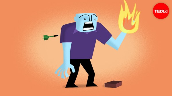
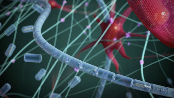
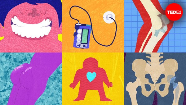
The largest organ in your body isn't your liver or your brain. It's your skin, with a surface area of about 20 square feet in adults. Though different areas of the skin have different characteristics, much of this surface performs similar functions, such as sweating, feeling heat and cold, and growing hair. But after a deep cut or wound, the newly healed skin will look different from the surrounding area, and may not fully regain all its abilities for a while, or at all. To understand why this happens, we need to look at the structure of the human skin. The top layer, called the epidermis, consists mostly of hardened cells, called keratinocytes, and provides protection. Since its outer layer is constantly being shed and renewed, it's pretty easy to repair. But sometimes a wound penetrates into the dermis, which contains blood vessels and the various glands and nerve endings that enable the skin's many functions. And when that happens, it triggers the four overlapping stages of the regenerative process. The first stage, hemostasis, is the skin's response to two immediate threats: that you're now losing blood and that the physical barrier of the epidermis has been compromised. As the blood vessels tighten to minimize the bleeding, in a process known as vasoconstriction, both threats are averted by forming a blood clot. A special protein known as fibrin forms cross-links on the top of the skin, preventing blood from flowing out and bacteria or pathogens from getting in. After about three hours of this, the skin begins to turn red, signaling the next stage, inflammation. With bleeding under control and the barrier secured, the body sends special cells to fight any pathogens that may have gotten through. Among the most important of these are white blood cells, known as macrophages, which devour bacteria and damage tissue through a process known as phagocytosis, in addition to producing growth factors to spur healing. And because these tiny soldiers need to travel through the blood to get to the wound site, the previously constricted blood vessels now expand in a process called vasodilation. About two to three days after the wound, the proliferative stage occurs, when fibroblast cells begin to enter the wound. In the process of collagen deposition, they produce a fibrous protein called collagen in the wound site, forming connective skin tissue to replace the fibrin from before. As epidermal cells divide to reform the outer layer of skin, the dermis contracts to close the wound. Finally, in the fourth stage of remodeling, the wound matures as the newly deposited collagen is rearranged and converted into specific types. Through this process, which can take over a year, the tensile strength of the new skin is improved, and blood vessels and other connections are strengthened. With time, the new tissue can reach from 50-80% of some of its original healthy function, depending on the severity of the initial wound and on the function itself. But because the skin does not fully recover, scarring continues to be a major clinical issue for doctors around the world. And even though researchers have made significant strides in understanding the healing process, many fundamental mysteries remain unresolved. For instance, do fibroblast cells arrive from the blood vessels or from skin tissue adjacent to the wound? And why do some other mammals, such as deer, heal their wounds much more efficiently and completely than humans? By finding the answers to these questions and others, we may one day be able to heal ourselves so well that scars will be just a memory.
Cel mai mare organ al corpului nu e ficatul sau creierul, ci pielea, cu o suprafață de circa 1,86 m² la adulți. Deși diferite zone ale pielii au trăsături diferite, majoritatea acestei suprafețe îndeplinește funcții similare, cum ar fi transpirația, percepția căldurii și a frigului și creșterea părului. Însă după o tăietură sau rană adâncă, pielea recent cicatrizată va arăta diferit față de suprafața dimprejurul său și s-ar putea să nu-și recapete complet toate funcțiile ceva timp sau chiar deloc. Pentru a înțelege această cauză, trebuie să analizăm structura pielii. Stratul superficial, numit epidermă, constă în mare parte din celule întărite, numite keratinocite, și asigură protecție. Deoarece stratul său exterior se schimbă și se reînnoiește treptat, este destul de ușor de vindecat. Uneori, însă, rana pătrunde derma ce conține vase sangvine, diferite glande și terminații nervoase ce asigură îndeplinirea diverselor funcții ale pielii. Când are loc acest lucru, se declanșează patru etape succesive ale procesului de regenerare. Prima etapă, hemostaza, e reacția pielii la două amenințări iminente: că pierzi sânge și că bariera fizică a epidermei a fost afectată. Odată ce vasele sangvine se contractă pentru a reduce sângerarea, proces numit vasoconstricție, ambele amenințări sunt prevenite prin formarea unui cheag de sânge. O proteină specială, numită fibrină, formează legături transversale la suprafața pielii, împiedicând sângerarea și pătrunderea bacteriilor sau a agenților patogeni. După circa trei ore de la acest proces, pielea începe să se înroșească, indicând următoarea etapă, cea a inflamației. Odată ce sângerarea a fost oprită iar bariera securizată, organismul generează celule pentru a lupta cu posibilii agenți patogeni pătrunși. Printre cele mai importante celule se numără leucocitele, cunoscute sub denumirea de macrofage, ce devorează bacteriile și distrug țesuturile printr-un proces numit fagocitoză, producând și factori de creștere pentru a stimula vindecarea. Întrucât acești mici „soldați” trebuie să circule prin sânge pentru a ajunge la locul rănii, vasele de sânge contractate anterior se dilată acum, printr-un proces numit vasodilatație. După aproximativ 2-3 zile de la apariția rănii, are loc etapa proliferativă, când celulele fibroblaste încep să pătrundă în rană. În procesul de depunere a colagenului, acestea încep să producă în zona rănii o proteină fibroasă, numită colagen, formând țesutul conjunctiv al pielii, pentru a înlocui fibrina de dinainte. Cât timp celulele epidermice se divizează pentru a reface stratul superficial, derma se contractă pentru a închide rana. În final, în a patra etapă, numită remodelare, rana se vindecă pe măsură ce colagenul recent depozitat e rearanjat și transformat în diferite tipuri. În cadrul acestui proces, care poate dura mai mult de un an, e îmbunătățită rezistența la întindere a pielii nou formate, iar vasele sangvine și alte structuri sunt întărite. Cu timpul, țesutul nou format poate dobândi între 50% și 80% din funcția sa sănătoasă inițială, în funcție de gravitatea rănii suferite și de funcție în sine. Însă întrucât pielea nu se recuperează complet, cicatrizarea rămâne o problemă clinică majoră pentru medicii din întreaga lume. Cu toate că cercetătorii au realizat progrese semnificative în înțelegerea procesului de vindecare, multe dintre misterele esențiale rămân nerezolvate. De exemplu, celule fibroblaste provin de la vasele de sânge sau de la țesutul cutanat adiacent rănii? Și de ce la unele mamifere, cum ar fi căprioara, rănile se vindecă mult mai rapid și complet decât la oameni? Când vom găsi răspuns la aceste întrebări, precum și la altele, vom putea într-o bună zi să ne tratăm atât de bine