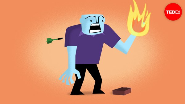
The largest organ in your body isn't your liver or your brain. It's your skin, with a surface area of about 20 square feet in adults. Though different areas of the skin have different characteristics, much of this surface performs similar functions, such as sweating, feeling heat and cold, and growing hair. But after a deep cut or wound, the newly healed skin will look different from the surrounding area, and may not fully regain all its abilities for a while, or at all. To understand why this happens, we need to look at the structure of the human skin. The top layer, called the epidermis, consists mostly of hardened cells, called keratinocytes, and provides protection. Since its outer layer is constantly being shed and renewed, it's pretty easy to repair. But sometimes a wound penetrates into the dermis, which contains blood vessels and the various glands and nerve endings that enable the skin's many functions. And when that happens, it triggers the four overlapping stages of the regenerative process. The first stage, hemostasis, is the skin's response to two immediate threats: that you're now losing blood and that the physical barrier of the epidermis has been compromised. As the blood vessels tighten to minimize the bleeding, in a process known as vasoconstriction, both threats are averted by forming a blood clot. A special protein known as fibrin forms cross-links on the top of the skin, preventing blood from flowing out and bacteria or pathogens from getting in. After about three hours of this, the skin begins to turn red, signaling the next stage, inflammation. With bleeding under control and the barrier secured, the body sends special cells to fight any pathogens that may have gotten through. Among the most important of these are white blood cells, known as macrophages, which devour bacteria and damage tissue through a process known as phagocytosis, in addition to producing growth factors to spur healing. And because these tiny soldiers need to travel through the blood to get to the wound site, the previously constricted blood vessels now expand in a process called vasodilation. About two to three days after the wound, the proliferative stage occurs, when fibroblast cells begin to enter the wound. In the process of collagen deposition, they produce a fibrous protein called collagen in the wound site, forming connective skin tissue to replace the fibrin from before. As epidermal cells divide to reform the outer layer of skin, the dermis contracts to close the wound. Finally, in the fourth stage of remodeling, the wound matures as the newly deposited collagen is rearranged and converted into specific types. Through this process, which can take over a year, the tensile strength of the new skin is improved, and blood vessels and other connections are strengthened. With time, the new tissue can reach from 50-80% of some of its original healthy function, depending on the severity of the initial wound and on the function itself. But because the skin does not fully recover, scarring continues to be a major clinical issue for doctors around the world. And even though researchers have made significant strides in understanding the healing process, many fundamental mysteries remain unresolved. For instance, do fibroblast cells arrive from the blood vessels or from skin tissue adjacent to the wound? And why do some other mammals, such as deer, heal their wounds much more efficiently and completely than humans? By finding the answers to these questions and others, we may one day be able to heal ourselves so well that scars will be just a memory.
El órgano más grande de tu cuerpo no es tu hígado o tu cerebro. Es tu piel, con una superficie de unos 1,86 metros cuadrados en los adultos. Aunque muchas áreas de la piel tienen características diferentes, mucha de esta superficie realiza funciones similares, como transpirar, sentir calor y frío, y crecer pelo. Pero luego de un corte profundo o una herida, la nueva piel se verá diferente al área que la rodea, y puede que no recupere totalmente sus habilidades durante un tiempo, o en absoluto. Para comprender por qué sucede esto, necesitamos mirar la estructura de la piel humana. La capa superior, llamada epidermis, consiste sobretodo en células endurecidas, llamadas queratinocitos, que dan protección. Dado que esta capa externa se cambia y renueva constantemente, es muy fácil repararla. Pero a veces una herida penetra en la dermis, la cual contiene vasos sanguíneos, varias glándulas y terminaciones nerviosas que permiten las muchas funciones de la piel. Y cuando eso sucede, desencadena los 4 procesos sobrepuestos del proceso de regeneración. La primera etapa, hemostasia, es la respuesta de la piel a dos amenazas inmediatas: que estás perdiendo sangre y que la barrera física de la epidermis ha sido dañada. A medida que los vasos sanguíneos se contrajeron para reducir al mínimo el sangrado, en un proceso llamado vasoconstricción, las dos amenazas se evitan al formar un coágulo de sangre. Una proteína especial conocida como fibrina, forma enlaces cruzados en la superficie de la piel, evitando que la sangre fluya hacia afuera y que entren bacterias o patógenos. Luego de alrededor de 3 horas, la piel se empieza a poner roja, indicando la nueva etapa, la inflamación. Cuando el sangrado está bajo control y la barrera asegurada, el cuerpo envía células especializadas para luchar contra cualquier patógeno que pudiera haber entrado. Entre las más importantes de estas están los glóbulos blancos, llamados macrófagos, que devoran a bacterias y tejido dañado en un proceso llamado fagocitosis, además de producir factores de crecimiento para estimular la curación. Y dado que estos diminutos soldados necesitan viajar por la sangre para llegar hasta el sitio dañado, los vasos sanguíneos contraídos, ahora se expanden en un proceso llamado vasodilatación. Dos o tres días después de la herida, ocurre la etapa proliferativa, en que las células fibroblastos entran en la herida. En el proceso de deposición de colágeno, producen una proteína fibrosa llamada colágeno en la zona herida, formando tejido conectivo para reemplazar la fibrina anterior. Mientras que las células epidérmicas se dividen para regenerar la capa externa de la piel, la dermis se contrae para cerrar la herida. Finalmente, en la cuarta etapa de remodelación, la herida madura cuando el colágeno depositado se reorganiza y se convierte en tipos específicos. Mediante este proceso, que puede tomar hasta un año, la resistencia a la tracción de la piel nueva mejora, y los vasos sanguíneos y otras conexiones se fortalecen. Con el tiempo, el nuevo tejido puede alcanzar el 50 a 80% de sus funciones originales, dependiendo de la gravedad de la herida inicial y de la función en sí misma. Pero como la piel no se recupera 100%, la cicatrización sigue siendo un problema clínico importante para los médicos de todo el mundo. Y a pesar de que los investigadores han hecho avances significativos en la comprensión del proceso de curación, muchos misterios fundamentales permanecen sin resolver. Por ejemplo, ¿los fibroblastos llegan por los vasos sanguíneos o del tejido de la piel adyacente a la herida? ¿Y por qué algunos otros mamíferos, como los ciervos, curan sus heridas mucho más eficiente y completamente que los seres humanos? Encontrando las respuestas a estas y otras preguntas, podremos ser capaces, algún día, de sanarnos tan bien a nosotros mismos