In the space that used to house one transistor, we can now fit one billion. That made it so that a computer the size of an entire room now fits in your pocket. You might say the future is small.
예전에 트랜지스터 하나가 차지하던 공간에 지금은 10억 개를 넣을 수 있습니다. 방 하나만큼 크던 컴퓨터가 주머니에 들어갈 만큼 작아졌지요. 미래의 세상은 작다고 할 수도 있겠죠.
As an engineer, I'm inspired by this miniaturization revolution in computers. As a physician, I wonder whether we could use it to reduce the number of lives lost due to one of the fastest-growing diseases on Earth: cancer. Now when I say that, what most people hear me say is that we're working on curing cancer. And we are. But it turns out that there's an incredible opportunity to save lives through the early detection and prevention of cancer.
엔지니어로서 저는 컴퓨터가 급격히 소형화된 점이 인상깊었습니다. 내과 의사로서 급증하는 질병으로 목숨을 잃는 환자를 줄이는데 이 기술을 활용하면 어떨까 생각했습니다. 암. 제가 암이라고 하면 다들 암을 치료하는 일을 말하는 걸로 알죠. 그렇긴 합니다. 하지만 암을 조기에 발견하고 예방하면 생명을 살릴 기회는 엄청나게 높아집니다.
Worldwide, over two-thirds of deaths due to cancer are fully preventable using methods that we already have in hand today. Things like vaccination, timely screening and of course, stopping smoking. But even with the best tools and technologies that we have today, some tumors can't be detected until 10 years after they've started growing, when they are 50 million cancer cells strong. What if we had better technologies to detect some of these more deadly cancers sooner, when they could be removed, when they were just getting started?
암 사망의 2/3 이상은 요즘 기술로도 충분히 막을 수 있습니다. 백신이나 적시 검진같은 것들이죠. 물론 금연도 포함되구요. 하지만 최고의 방법과 기술을 써도 어떤 종양은 10년간 자라나 5천만 개의 암세포로 강해지고 나서야 발견됩니다. 만약 우리가 이런 치명적인 암을 더 일찍 발견할 수 있다면 어떨까요? 암이 발생하기 시작하고 이를 제거할 수 있을 때 말입니다.
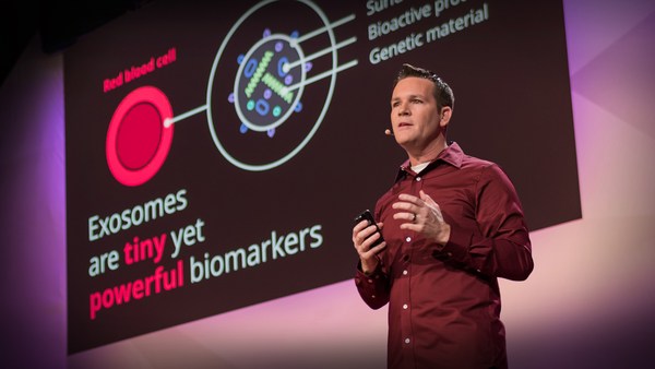
Let me tell you about how miniaturization might get us there. This is a microscope in a typical lab that a pathologist would use for looking at a tissue specimen, like a biopsy or a pap smear. This $7,000 microscope would be used by somebody with years of specialized training to spot cancer cells. This is an image from a colleague of mine at Rice University, Rebecca Richards-Kortum. What she and her team have done is miniaturize that whole microscope into this $10 part, and it fits on the end of an optical fiber. Now what that means is instead of taking a sample from a patient and sending it to the microscope, you can bring the microscope to the patient. And then, instead of requiring a specialist to look at the images, you can train the computer to score normal versus cancerous cells.
소형화 기술이 여기에 어떻게 사용되는지 말씀드리겠습니다. 이건 보통 연구실에 있는 현미경인데요. 병리학자가 생검이나 팹 테스트(자궁암 조기 검사)를 위해서 조직 표본을 살펴볼 때 쓰지요. 누군가는 수년간 전문 수련을 받고 700만원짜리 이 현미경으로 암세포를 찾을 겁니다. 이것은 라이스 대학의 제 동료인 레베카 리차드 코텀이 보내준 이미지입니다. 그 팀에서는 이 현미경 전체를 축소해서 광섬유 끝에 달기 좋은 만원 짜리 작은 부품으로 만들었습니다. 이제 환자에게서 샘플을 채취해 현미경으로 가져오는 대신에 현미경을 환자에게로 가져갈 수 있게 된거죠. 그리고 전문가가 이미지를 보는 대신에 컴퓨터를 훈련시켜 정상 세포와 암세포를 찾아보게 합니다.
Now this is important, because what they found working in rural communities, is that even when they have a mobile screening van that can go out into the community and perform exams and collect samples and send them to the central hospital for analysis, that days later, women get a call with an abnormal test result and they're asked to come in. Fully half of them don't turn up because they can't afford the trip. With the integrated microscope and computer analysis, Rebecca and her colleagues have been able to create a van that has both a diagnostic setup and a treatment setup. And what that means is that they can do a diagnosis and perform therapy on the spot, so no one is lost to follow up.
이 부분이 중요한거죠. 왜냐면 시골 마을에서는 이동 검사 차량으로 방문해 검사를 하고 샘플을 모아 병원 본부에 분석을 의뢰하러 보내고 며칠 후 환자는 비정상적인 검사 결과를 통보받고 병원으로 오라는 얘기를 듣죠. 그 환자들의 반은 형편이 안되서 병원을 방문하지도 못합니다. 현미경과 컴퓨터 분석이 통합되어 레베카와 동료들은 진단과 치료 장비를 갖춘 차량을 만들 수 있었습니다. 그래서 현장에서 진단하고 치료를 해서 후속 치료를 놓치는 사람이 없죠.
That's just one example of how miniaturization can save lives. Now as engineers, we think of this as straight-up miniaturization. You took a big thing and you made it little. But what I told you before about computers was that they transformed our lives when they became small enough for us to take them everywhere. So what is the transformational equivalent like that in medicine? Well, what if you had a detector that was so small that it could circulate in your body, find the tumor all by itself and send a signal to the outside world? It sounds a little bit like science fiction. But actually, nanotechnology allows us to do just that. Nanotechnology allows us to shrink the parts that make up the detector from the width of a human hair, which is 100 microns, to a thousand times smaller, which is 100 nanometers. And that has profound implications.
이것은 소형화 기술이 생명을 구하는 사례 중 하나일 뿐입니다. 이번에는 공학자로서 우리는 이런 것을 '그대로 소형화'라고 생각했습니다. 커다란 것을 작게 만들죠. 하지만 제가 앞서 컴퓨터를 어디로든 가져갈 정도로 충분히 작게 만들면 우리 생명도 구할 거라고 말씀드렸지요. 그러면 의약에서 이에 해당되는 것은 뭘까요? 자, 아주 작은 탐지기가 우리 몸을 돌아다니다가 스스로 종양을 발견하고 외부에 알려주면 어떨까요? 공상 과학처럼 들리시죠. 하지만 사실 나노기술로 이렇게 할 수 있습니다. 나노 기술로 탐지기를 사람 머리카락의 두께만큼 줄이면 100 마이크론이 되는데 이걸 천 배 더 줄이면 100 나노미터가 됩니다. 그러면 엄청난 일이 가능해져요.
It turns out that materials actually change their properties at the nanoscale. You take a common material like gold, and you grind it into dust, into gold nanoparticles, and it changes from looking gold to looking red. If you take a more exotic material like cadmium selenide -- forms a big, black crystal -- if you make nanocrystals out of this material and you put it in a liquid, and you shine light on it, they glow. And they glow blue, green, yellow, orange, red, depending only on their size. It's wild! Can you imagine an object like that in the macro world? It would be like all the denim jeans in your closet are all made of cotton, but they are different colors depending only on their size.
물질은 나노 단위가 되면 속성이 바뀝니다. 금을 예로 들면 먼지로 갈아서 금 나노 입자로 만들면 금으로 보이지 않고 붉게 보이지요. 셀렌화 카드뮴같은 좀 더 낯선 물질을 예로 들어볼까요. 이 물질은 원래 큰 검은 결정을 형성하는데 나노 결정으로 만들어 액체에 넣으면 빛나는 불빛을 얻을 수 있어요. 크기에 따라 파랑, 초록, 노랑, 주황, 빨강으로 변합니다. 말도 안되죠! 매크로 세상에서 이런 물체를 상상할 수 있나요? 내 옷장의 청바지가 모두 면 소재인데도 크기만 다르면 색상도 달라지는 것과 같다구요.
(Laughter)
(웃음)
So as a physician, what's just as interesting to me is that it's not just the color of materials that changes at the nanoscale; the way they travel in your body also changes. And this is the kind of observation that we're going to use to make a better cancer detector.
그래서 의사로서 저는 나노 단위의 크기에 따라 물질의 색상이 달라지는 것 만큼이나 몸 속을 돌아다니는 방식도 달라진다는 점이 흥미로왔습니다. 이런 관찰 결과를 이용해 더 나은 암 탐지기를 만들 것입니다.
So let me show you what I mean. This is a blood vessel in the body. Surrounding the blood vessel is a tumor. We're going to inject nanoparticles into the blood vessel and watch how they travel from the bloodstream into the tumor. Now it turns out that the blood vessels of many tumors are leaky, and so nanoparticles can leak out from the bloodstream into the tumor. Whether they leak out depends on their size. So in this image, the smaller, hundred-nanometer, blue nanoparticles are leaking out, and the larger, 500-nanometer, red nanoparticles are stuck in the bloodstream. So that means as an engineer, depending on how big or small I make a material, I can change where it goes in your body.
제가 말씀드리는 내용을 보여드리겠습니다. 이건 몸 속의 혈관입니다. 혈관을 종양이 감싸고 있지요. 나노 입자를 혈관에 주입하면 혈류를 타고 어떻게 종양으로 가는지 보겠습니다. 종양의 혈관에는 새는 구멍이 있는데 혈류에 있던 나노 입자는 이 구멍으로 빠져나와 종양으로 갑니다. 나노입자의 크기에 따라 빠져나올지 말지 결정되죠. 이 영상에서 파랑색인 100 나노미터짜리 작은 나노 입자가 새어 나오고 있고 빨간색인 500 나노미터짜리 큰 나노 입자는 혈류에 남아 있지요. 공학자의 눈으로 보니 입자를 어떤 크기로 만드느냐에 따라 몸의 어디로 보낼지 정할 수 있겠더라구요.
In my lab, we recently made a cancer nanodetector that is so small that it could travel into the body and look for tumors. We designed it to listen for tumor invasion: the orchestra of chemical signals that tumors need to make to spread. For a tumor to break out of the tissue that it's born in, it has to make chemicals called enzymes to chew through the scaffolding of tissues. We designed these nanoparticles to be activated by these enzymes. One enzyme can activate a thousand of these chemical reactions in an hour. Now in engineering, we call that one-to-a-thousand ratio a form of amplification, and it makes something ultrasensitive. So we've made an ultrasensitive cancer detector.
우리 연구실에서 최근에 나노 암 탐지기를 만들었는데 아주 작아서 몸 속을 다니며 종양을 찾아줍니다. 종양이 증식하는지를 감지하도록 설계했지요. 다시 말해, 종양이 증식될 때 발생되는 화학 신호들을 알아차립니다. 종양이 발생한 조직에서 밖으로 커져 나오려면 근육의 기반이 되는 부분을 잘게 뜯어서 나와야 하는데 이 때 효소라고 하는 화학 물질을 만들어 냅니다. 이 효소가 우리가 만든 나노 입자를 가동시키게 만든거지요. 효소 하나로 이런 화학 반응 천 개를 활성화하는데 한 시간이 걸리지 않습니다. 공학에서는 이렇게 1 대 1000로 반응하는 것을 증폭이라고 하는데 초고감도 장비를 만들 수 있습니다. 우리는 초고감도 암 탐지기를 만들었구요.
OK, but how do I get this activated signal to the outside world, where I can act on it? For this, we're going to use one more piece of nanoscale biology, and that has to do with the kidney. The kidney is a filter. Its job is to filter out the blood and put waste into the urine. It turns out that what the kidney filters is also dependent on size. So in this image, what you can see is that everything smaller than five nanometers is going from the blood, through the kidney, into the urine, and everything else that's bigger is retained. OK, so if I make a 100-nanometer cancer detector, I inject it in the bloodstream, it can leak into the tumor where it's activated by tumor enzymes to release a small signal that is small enough to be filtered out of the kidney and put into the urine, I have a signal in the outside world that I can detect.
자 그런데 이런 신호를 어떻게 외부로 보내 제가 조치할 수 있을까요? 우리는 콩팥과 관련한 나노 생물학 기술을 이용할 겁니다. 콩팥은 필터죠. 혈액을 거르고 남은 찌꺼기를 소변으로 보냅니다. 콩팥은 물질의 크기를 기준으로 입자를 걸러냅니다. 이 영상에서 보시듯이 혈액이 콩팥을 지나면 5 나노미터보다 작은 건 모두 소변으로 가고 그보다 큰 건 모두 남지요. 그러니까 제가 100 나노미터짜리 암 탐지기를 만들어 혈관에 주입하면 탐지기는 종양으로 침투해서 종양 효소에 의해 활성화되어 콩팥을 통과할 정도로 작은 물질을 방출하는 겁니다. 이 물질이 소변으로 나오면 저는 외부에서 그걸 감지하면 되겠죠.
OK, but there's one more problem. This is a tiny little signal, so how do I detect it? Well, the signal is just a molecule. They're molecules that we designed as engineers. They're completely synthetic, and we can design them so they are compatible with our tool of choice. If we want to use a really sensitive, fancy instrument called a mass spectrometer, then we make a molecule with a unique mass. Or maybe we want make something that's more inexpensive and portable. Then we make molecules that we can trap on paper, like a pregnancy test. In fact, there's a whole world of paper tests that are becoming available in a field called paper diagnostics.
그런데 한 가지 문제가 더 있어요. 이 신호 물질이 아주 작은데 제가 어떻게 감지하죠? 신호라고 해봐야 분자 하나일 뿐인데요. 공학자들이 만든 분자예요. 완전 합성 물질이니 우리 용도대로 만들 수 있지요. 질량 분석기라는 아주 멋지고 똑똑한 장비를 쓴다면 질량이 독특한 분자를 만드는 겁니다. 아니면 더 저렴하고 휴대할 만한 것도 좋겠지요. 그러면 분자를 만들어서 임신 테스트기같은 종이로 감지할 수 있습니다. 사실 진단 분야에 온갖 종류의 진단지 테스트가 나오고 있습니다.
Alright, where are we going with this? What I'm going to tell you next, as a lifelong researcher, represents a dream of mine. I can't say that's it's a promise; it's a dream. But I think we all have to have dreams to keep us pushing forward, even -- and maybe especially -- cancer researchers.
자, 우리는 이걸로 뭘 할까요? 제가 다음에 말씀드릴 것은 제가 평생 연구하려는 꿈에 대한 것입니다. 약속이라고 말씀드릴 수는 없고 꿈이라고 하겠습니다. 우리는 앞으로 나가기 위해 지향할 꿈이 필요합니다 암 연구자일지라도요. 어쩌면 암 연구자니까 더 그래야하죠.
I'm going to tell you what I hope will happen with my technology, that my team and I will put our hearts and souls into making a reality. OK, here goes. I dream that one day, instead of going into an expensive screening facility to get a colonoscopy, or a mammogram, or a pap smear, that you could get a shot, wait an hour, and do a urine test on a paper strip. I imagine that this could even happen without the need for steady electricity, or a medical professional in the room. Maybe they could be far away and connected only by the image on a smartphone.
제가 말씀드리려는 것은 이 기술로 제가 바라는 것을 만들어 저와 팀이 심혈을 기울여서 실현해 내겠다는 것입니다. 그럼, 시작해보죠. 언젠가는 고가의 장비를 이용해서 결장 내시술, 유방 X선 촬영 검사, 팹 검사를 하는 대신 주사를 한 대 맞고 한 시간 기다린 후 종이에 소변 검사를 하게 될 거예요. 제 생각에는 지속적인 전력 공급이 없이도 의학 전문가가 현장에 없이도 이렇게 될 수 있어요. 전문가는 멀리서 스마트폰 영상으로 볼 수 있겠지요.
Now I know this sounds like a dream, but in the lab we already have this working in mice, where it works better than existing methods for the detection of lung, colon and ovarian cancer. And I hope that what this means is that one day we can detect tumors in patients sooner than 10 years after they've started growing, in all walks of life, all around the globe, and that this would lead to earlier treatments, and that we could save more lives than we can today, with early detection.
허황되게 들리겠지만 우리 연구실에서 이미 쥐를 대상으로 실험하고 있고 폐암, 대장암이나 난소암을 발견하는 데 기존 방법보다 탁월했습니다. 이 연구로 전 세계 각계 각층 환자에게서 종양이 발생한 지 10년이나 되기 전에 암을 검진하여 조기 치료로 오늘날보다 더 많은 생명을 살리는 날이 오기를 희망합니다.
Thank you.
감사합니다.
(Applause)
(박수)