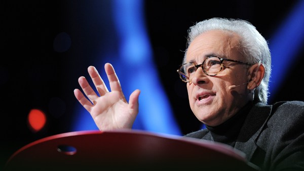
Today I want to tell you about a project being carried out by scientists all over the world to paint a neural portrait of the human mind. And the central idea of this work is that the human mind and brain is not a single, general-purpose processor, but a collection of highly specialized components, each solving a different specific problem, and yet collectively making up who we are as human beings and thinkers. To give you a feel for this idea,
Сьогодні я хочу розповісти про проект, який реалізується науковцями по всьому світу. Його ціллю є намалювати нейронний портрет людського розуму. І головна ідея цієї роботи в тому, що людський розум і мозок - це не є простий, багатоцільовий процесор, а колекція вузькоспеціалізованих деталей, кожна з яких вирішує іншу конкретну проблему, і при цьому вони роблять нас тими, ким ми є - людськими істотами та мислителями. Щоб ви могли краще зрозуміти цю ідею,
imagine the following scenario: You walk into your child's day care center. As usual, there's a dozen kids there waiting to get picked up, but this time, the children's faces look weirdly similar, and you can't figure out which child is yours. Do you need new glasses? Are you losing your mind? You run through a quick mental checklist. No, you seem to be thinking clearly, and your vision is perfectly sharp. And everything looks normal except the children's faces. You can see the faces, but they don't look distinctive, and none of them looks familiar, and it's only by spotting an orange hair ribbon that you find your daughter.
уявіть наступну ситуацію. Ви йдете до дитячого садку, куди ходить Ваша дитина. Зазвичай там є дванадцятеро дітей, які чекають, коли їх заберуть, але в цей раз дитячі обличчя виглядають незвично схожими, і Ви не можете з'ясувати, яка дитина Ваша. Вам потрібні нові окуляри? Чи Ви божеволієте? Ви швидко оцінюєте свій психічний стан. Та ні, Ви, здається, ясно мислите, і ваш зір показує гранично чітке зображення. І все виглядає нормально, окрім дитячих облич. Ви можете бачити обличчя, але їх не розпізнати, і жодне з них не виглядає добре знайомим. І лише виявивши помаранчеву стрічку на волоссі Ви знаходите свою дочку.
This sudden loss of the ability to recognize faces actually happens to people. It's called prosopagnosia, and it results from damage to a particular part of the brain. The striking thing about it is that only face recognition is impaired; everything else is just fine.
Така раптова втрата здатності розпізнавати обличчя справді відбувається з людьми. Це називається прозопагнозія, і є наслідком пошкодження певної частини мозку. Вражає те, що погіршується тільки розпізнавання облич; все інше працює просто відмінно.
Prosopagnosia is one of many surprisingly specific mental deficits that can happen after brain damage. These syndromes collectively have suggested for a long time that the mind is divvied up into distinct components, but the effort to discover those components has jumped to warp speed with the invention of brain imaging technology, especially MRI. So MRI enables you to see internal anatomy at high resolution, so I'm going to show you in a second a set of MRI cross-sectional images through a familiar object, and we're going to fly through them and you're going to try to figure out what the object is. Here we go.
Прозопагнозія є одним з багатьох на диво конкретних психологічних дефіцитів, який може відбутися після пошкодження головного мозку. Впродовж тривалого часу ці синдроми разом наводили на думку, що розум розділений на окремі компоненти, але спроба виявити ті компоненти досягла недосяжної швидкості з винаходом технології сканування головного мозку, особливо МРТ. МРТ дозволяє побачити внутрішню анатомію при високій роздільній здатності. Я збираюся показати вам набір МРТ зображень знайомих нам предметів у поперечному перерізі. Ми проглянемо їх наскрізь, і Ви спробуєте з'ясувати, які то є об'єкти. Приступаємо!
It's not that easy. It's an artichoke.
Це не так просто. Це артишок.
Okay, let's try another one, starting from the bottom and going through the top. Broccoli! It's a head of broccoli. Isn't it beautiful? I love that.
Добре, давайте спробуємо ще один. Починаємо з нижньої частини і переходимо вверх. Броколі! Це головка броколі. Хіба це не прекрасно? Мені подобається.
Okay, here's another one. It's a brain, of course. In fact, it's my brain. We're going through slices through my head like that. That's my nose over on the right, and now we're going over here, right there.
Добре, ось ще один. Це мозок, звісно. Насправді, це мій мозок. Ми проходимо через прошарки моєї голові. Це мій ніс справа. Зараз ми рухаємося сюди.
So this picture's nice, if I do say so myself, but it shows only anatomy. The really cool advance with functional imaging happened when scientists figured out how to make pictures that show not just anatomy but activity, that is, where neurons are firing. So here's how this works. Brains are like muscles. When they get active, they need increased blood flow to supply that activity, and lucky for us, blood flow control to the brain is local, so if a bunch of neurons, say, right there get active and start firing, then blood flow increases just right there. So functional MRI picks up on that blood flow increase, producing a higher MRI response where neural activity goes up.
Так ця картинка гарна, якщо я говорю так, але вона показує тільки анатомію. Дійсно здоровий прогрес у функціональному скануванні відбувся, коли вчені з'ясували, як зробити фотографії, які показують не тільки анатомію, але й діяльність, тобто, місце, де активуються нейрони. Ось, як це працює. Мозок, як м'язи. Коли вони стають активними, вони потребують збільшення припливу крові, щоб підтримувати цю діяльність, і на щастя для нас, управління кровотоку до мозку є локальним. Якщо в'язка нейронів, скажімо, прямо там стає активною і починає збуджуватися, потім кровотік збільшується прямо туди. Функціональна МРТ покращує збільшення кровотоку, виробляючи більш високу МРТ реакцію там, де нейронна активність зростає.
So to give you a concrete feel for how a functional MRI experiment goes and what you can learn from it and what you can't, let me describe one of the first studies I ever did. We wanted to know if there was a special part of the brain for recognizing faces, and there was already reason to think there might be such a thing based on this phenomenon of prosopagnosia that I described a moment ago, but nobody had ever seen that part of the brain in a normal person, so we set out to look for it. So I was the first subject. I went into the scanner, I lay on my back, I held my head as still as I could while staring at pictures of faces like these and objects like these and faces and objects for hours. So as somebody who has pretty close to the world record of total number of hours spent inside an MRI scanner, I can tell you that one of the skills that's really important for MRI research is bladder control. (Laughter)
Таким чином, щоб дати Вам конкретне розуміння про те, як функціональне МРТ дослідження триває і що Ви можете, і не можете дізнатися про нього, дозвольте мені описати одне з моїх перших досліджень. Ми хотіли знати, чи є спеціальна частина мозку для розпізнавання облич. У нас вже була причина думати, що саме така частина повинна існувати, базуючись на основі явища прозопагнозії, про яке я розповідала хвилину тому. Але ніхто ніколи не бачив цю частину мозку у нормальної людини, тому ми мали намір шукати її. Я була першою. Я підійшла до сканера, лягла на спину, і утримувала голову так нерухомо, як тільки могла, годинами розглядаючи фотографії облич, таких як ці, і таких предметів, як ці. І обличчя, і предмети годинами. Наблизившись дуже близько до світового рекорду загальної кількості годин, проведених всередині МРТ-сканера, я можу вам сказати, що одна з навичок, яка дуже важлива для МРТ дослідження, - це контроль сечового міхура. (Сміх)
When I got out of the scanner, I did a quick analysis of the data, looking for any parts of my brain that produced a higher response when I was looking at faces than when I was looking at objects, and here's what I saw. Now this image looks just awful by today's standards, but at the time I thought it was beautiful. What it shows is that region right there, that little blob, it's about the size of an olive and it's on the bottom surface of my brain about an inch straight in from right there. And what that part of my brain is doing is producing a higher MRI response, that is, higher neural activity, when I was looking at faces than when I was looking at objects. So that's pretty cool, but how do we know this isn't a fluke? Well, the easiest way is to just do the experiment again. So I got back in the scanner, I looked at more faces and I looked at more objects and I got a similar blob, and then I did it again and I did it again and again and again, and around about then I decided to believe it was for real. But still, maybe this is something weird about my brain and no one else has one of these things in there, so to find out, we scanned a bunch of other people and found that pretty much everyone has that little face-processing region in a similar neighborhood of the brain.
Коли я вийшла з-під сканера, я зробила швидкий аналіз даних, шукаючи певні частини мого мозку, які продукували сильнішу реакцію, коли я дивилася на обличчя, ніж тоді, коли я дивилася на предмети. І ось що я побачила. Зараз це зображення виглядає просто жахливо, за сьогоднішніми мірками, але в той час я думала, що воно було красивим. На ньому показана ділянка, та мала крапля, розміром з маслину, яка розташована в нижній частині мого мозку близько двох з половиною сантиметрів праворуч. Ця частина мого мозку дає вищу МРТ відповідь. Тобто вищу нейронну активність, коли я дивилася на обличчя, ніж тоді, коли я дивилася на предмети. Що ж, це дуже класно, але як ми знатимемо, що це не випадковість? Найпростіший спосіб - це повторити експеримент ще раз. Тому я повернулася під сканер, я продивилася більшу кількість облич та предметів і отримала аналогічну грудочку, а потім я зробила це ще раз, і ще раз, і ще, і ще. І десь після того я повірила, що це дійсно так. Але все-таки, може, це щось дивне з моїм мозком і ніхто інший не має нічого такого? Щоб з'ясувати це, ми просканували групу людей, і виявили, що в значній мірі кожен має ту маленьку ділянку для обробки облич в аналогічній частині мозку.
So the next question was, what does this thing really do? Is it really specialized just for face recognition? Well, maybe not, right? Maybe it responds not only to faces but to any body part. Maybe it responds to anything human or anything alive or anything round. The only way to be really sure that that region is specialized for face recognition is to rule out all of those hypotheses. So we spent much of the next couple of years scanning subjects while they looked at lots of different kinds of images, and we showed that that part of the brain responds strongly when you look at any images that are faces of any kind, and it responds much less strongly to any image you show that isn't a face, like some of these.
Тому наступним запитанням було те, що робить ця частина насправді? Чи дійсно вона спеціалізується тільки на розпізнаванні облич? Можливо й ні, погодьтеся? Можливо, вона відповідає не тільки за обличчя, але й за будь-яку частину тіла? Можливо, вона реагує на все людське чи щось живе, чи щось кругле? Єдиний спосіб впевнитися, що ця ділянка спеціалізується на розпізнаванні обличчя, - це перевірити решту гіпотез. Найближчі кілька років ми провели багато часу, скануючи людей, коли вони дивилися на безліч різноманітних видів зображень, і ми встановили, що та частина мозку реагує сильніше, коли Ви дивитеся на зображення облич будь-якого типу. Вона реагує значно слабше на зображення, на решту зображень, як, наприклад, ці.
So have we finally nailed the case that this region is necessary for face recognition? No, we haven't. Brain imaging can never tell you if a region is necessary for anything. All you can do with brain imaging is watch regions turn on and off as people think different thoughts. To tell if a part of the brain is necessary for a mental function, you need to mess with it and see what happens, and normally we don't get to do that. But an amazing opportunity came about very recently when a couple of colleagues of mine tested this man who has epilepsy and who is shown here in his hospital bed where he's just had electrodes placed on the surface of his brain to identify the source of his seizures. So it turned out by total chance that two of the electrodes happened to be right on top of his face area. So with the patient's consent, the doctors asked him what happened when they electrically stimulated that part of his brain. Now, the patient doesn't know where those electrodes are, and he's never heard of the face area. So let's watch what happens. It's going to start with a control condition that will say "Sham" nearly invisibly in red in the lower left, when no current is delivered, and you'll hear the neurologist speaking to the patient first. So let's watch.
Тож ми, нарешті, затвердили факт, що ця ділянка є необхідною для розпізнання обличчя? Ні. Сканування мозку ніколи не скаже Вам, чи є ця область необхідною для чого-небудь. Все, що Ви можете зробити із зображенням головного мозку - це дивитися, як ділянка вмикається і вимикається, коли люди думають про щось. Щоб дізнатися, чи необхідна якась частина мозку для розумової діяльності, Вам треба поморочитися з цим і подивитися, що відбувається, і, як правило, ми не робимо цього. Але зовсім недавно трапилася дивовижна можливість, коли кілька моїх колег перевіряли цього чоловіка, який страждає на епілепсію. Тут він на лікарняному ліжку, де йому щойно розмістили електроди на поверхні мозку, щоб визначити джерело його припадків. Цілком випадково виявилося, що два електроди опинилися над ділянкою розпізнавання облич. За згодою пацієнта, лікарі запитали в нього, що відбувалося, коли вони електрично стимулювали ту частину його мозку. Пацієнт не знає, де ці електроди розміщені, і він ніколи не чув про ділянку розпізнавання облич. Отже, давайте подивимося, що відбувалося. Все починається з умови контролю, коли буде майже непомітний червоний напис "SHAM" в лівому нижньому кутку, коли струм не подається. Ви почуєте спершу розмову невролога з пацієнтом. Давайте дивитися.
(Video) Neurologist: Okay, just look at my face and tell me what happens when I do this. All right?
(Відео) Невролог: Добре, просто подивіться на моє обличчя і скажіть мені, що відбувається, коли я роблю так. Добре?
Patient: Okay.
Пацієнт: Добре.
Neurologist: One, two, three.
Невролог: Один, два, три.
Patient: Nothing. Neurologist: Nothing? Okay. I'm going to do it one more time. Look at my face. One, two, three.
Пацієнт: Нічого. Невролог: Нічого? Добре. Я зроблю це ще раз. Подивіться на моє обличчя. Один, два, три.
Patient: You just turned into somebody else. Your face metamorphosed. Your nose got saggy, it went to the left. You almost looked like somebody I'd seen before, but somebody different. That was a trip. (Laughter)
Пацієнт: Ви просто перетворилися на когось іншого. Ваше обличчя видозмінилося. Ваш ніс провис, він схилився вліво. Ви майже схожі на когось, кого я бачив раніше, але когось іншого. Оце була подорож. (Сміх)
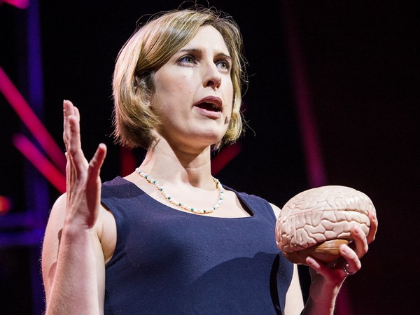
Nancy Kanwisher: So this experiment — (Applause) — this experiment finally nails the case that this region of the brain is not only selectively responsive to faces but causally involved in face perception. So I went through all of these details about the face region to show you what it takes to really establish that a part of the brain is selectively involved in a specific mental process. Next, I'll go through much more quickly some of the other specialized regions of the brain that we and others have found. So to do this, I've spent a lot of time in the scanner over the last month so I can show you these things in my brain.
Ненсі Кенвішер: Цей експеримент - (Оплески) - Цей експеримент, нарешті, підтверджує аргумент, що ця ділянка мозку не тільки вибірково реагує на обличчя, але й принагідно бере участь у сприйнятті обличчя. Я пройшла через усі ці деталі про ділянки обличчя, щоб показати вам, як довелося встановлювати той факт, що частина мозку селективно бере участь у конкретному психічному процесі. Далі я швидко розповім про інші спеціалізовані ділянки мозку, які ми та інші науковці знайшли. Щоб здійснити це, я провела багато часу під сканером за останній місяць. Я можу показати Вам ці речі в моєму мозку.
So let's get started. Here's my right hemisphere. So we're oriented like that. You're looking at my head this way. Imagine taking the skull off and looking at the surface of the brain like that. Okay, now as you can see, the surface of the brain is all folded up. So that's not good. Stuff could be hidden in there. We want to see the whole thing, so let's inflate it so we can see the whole thing. Next, let's find that face area I've been talking about that responds to images like these. To see that, let's turn the brain around and look on the inside surface on the bottom, and there it is, that's my face area. Just to the right of that is another region that is shown in purple that responds when you process color information, and near those regions are other regions that are involved in perceiving places, like right now, I'm seeing this layout of space around me and these regions in green right there are really active. There's another one out on the outside surface again where there's a couple more face regions as well. Also in this vicinity is a region that's selectively involved in processing visual motion, like these moving dots here, and that's in yellow at the bottom of the brain, and near that is a region that responds when you look at images of bodies and body parts like these, and that region is shown in lime green at the bottom of the brain.
Так що, давайте розпочнемо. Ось моя права півкуля. Зображення орієнтоване так. Ви дивитеся на мою голову в такий спосіб. Уявіть, що забираємо череп і дивимося на поверхню мозку ось так. Як Ви бачите, поверхня мозку вся в складках. Це не зручно. Матеріал може бути прихований там. Ми хочемо побачити цілісну річ, так що давайте розпрямимо її, щоб ми могли бачити все це. Давайте знайдемо ділянку розпізнавання облич, яка реагує на ось такі зображення. Щоб побачити це, давайте повернемо мозок навколо і побачимо внутрішню поверхню знизу. Ось вона, ця ділянка. Праворуч є ще одна ділянка, яка показана фіолетовим кольором, вона відповідає за обробку кольорової інформації. Біля тих та інших ділянок, які беруть участь у сприйнятті місця. Я бачу цю схему простору навколо себе і ці зелені ділянки є дійсно активними. Є ще одна на зовнішній поверхні, де є дві додаткові ділянки розпізнавання облич. Крім того поблизу є ділянка, яка селективно бере участь в обробці візуального руху, як оці рухомі точки. Вона позначена жовтим кольором у нижній частині головного мозку. Поруч є ділянка, яка реагує на зображення тіла і частин тіла, як це. Та ділянка зафарбована жовто-зеленим в нижній частині головного мозку.
Now all these regions I've shown you so far are involved in specific aspects of visual perception. Do we also have specialized brain regions for other senses, like hearing? Yes, we do. So if we turn the brain around a little bit, here's a region in dark blue that we reported just a couple of months ago, and this region responds strongly when you hear sounds with pitch, like these. (Sirens) (Cello music) (Doorbell) In contrast, that same region does not respond strongly when you hear perfectly familiar sounds that don't have a clear pitch, like these. (Chomping) (Drum roll) (Toilet flushing)
Зараз всі ці ділянки, які я показала Вам щойно, беруть участь у конкретних аспектах візуального сприйняття. Чи є у нас спеціалізовані ділянки мозку, для інших відчуттів, таких як слух? Так, є. Якщо ми трохи повернемо мозок навколо, ось область темно-синього кольору, про яку ми повідомили пару місяців тому. Ця ділянка сильно реагує, коли ви чуєте звуки з високим тоном, як ось ці. (Звук сирени) (Звуки віолончелі) (Дверний дзвінок) На відміну від цього, та ділянка не реагує сильно, коли ви чуєте добре знайомі звуки, які не мають високого тону. (Жування) (Барабанний дріб) (Змив у туалеті)
Okay. Next to the pitch region is another set of regions that are selectively responsive when you hear the sounds of speech.
Поруч з ділянкою "високого тону" є інша група ділянок, які вибірково реагують на звуки мови.
Okay, now let's look at these same regions. In my left hemisphere, there's a similar arrangement — not identical, but similar — and most of the same regions are in here, albeit sometimes different in size.
Тепер давайте подивимося на такі ж ділянки. У моїй лівій півкулі є аналогічний пристрій - не ідентичний, але схожий. Більшість з тих же ділянок є тут, хоча іноді відрізняються розміром.
Now, everything I've shown you so far are regions that are involved in different aspects of perception, vision and hearing. Do we also have specialized brain regions for really fancy, complicated mental processes? Yes, we do. So here in pink are my language regions. So it's been known for a very long time that that general vicinity of the brain is involved in processing language, but we showed very recently that these pink regions respond extremely selectively. They respond when you understand the meaning of a sentence, but not when you do other complex mental things, like mental arithmetic or holding information in memory or appreciating the complex structure in a piece of music.
Все, що я показувала Вам досі, - це ділянки, які беруть участь у різних аспектах сприйняття, зору та слуху. Чи маємо ми також спеціалізовані ділянки мозку для дійсно химерних та складних психічних процесів? Так, маємо. Ось рожевим зафарбовані мої "мовні" ділянки. Протягом довгого часу було відомо, що та ділянка мозку бере участь в обробці мови, але зовсім недавно ми показали, що ці рожеві ділянки реагують вкрай вибірково. Вони реагують, коли ви розумієте сенс речення, але не тоді, коли ви вирішуєте інші складні розумові завдання, як от усний рахунок, або запам'ятовування інформації, або оцінювання складної структури частини музичного твору.
The most amazing region that's been found yet is this one right here in turquoise. This region responds when you think about what another person is thinking. So that may seem crazy, but actually, we humans do this all the time. You're doing this when you realize that your partner is going to be worried if you don't call home to say you're running late. I'm doing this with that region of my brain right now when I realize that you guys are probably now wondering about all that gray, uncharted territory in the brain, and what's up with that?
Найдивовижніша ділянка, яка була знайдена, - це ось тут праворуч, бірюзового кольору. Ця ділянка реагує, коли ви задумуєтеся про те, що інша людина думає. Це може здатися божевіллям, але насправді, ми, люди, робимо це весь час. Ви робите це, коли ви розумієте, що ваш партнер турбуватиметься, якщо Ви не подзвоните і не попередите, що ви спізнюєтеся. Я роблю це за допомогою тієї ділянки мого мозку прямо зараз, коли я розумію, що ви, ймовірно, тепер зацікавилися всіма тими сірими незвіданими областями мозку, і тим, що там відбувається.
Well, I'm wondering about that too, and we're running a bunch of experiments in my lab right now to try to find a number of other possible specializations in the brain for other very specific mental functions. But importantly, I don't think we have specializations in the brain for every important mental function, even mental functions that may be critical for survival. In fact, a few years ago, there was a scientist in my lab who became quite convinced that he'd found a brain region for detecting food, and it responded really strongly in the scanner when people looked at images like this. And further, he found a similar response in more or less the same location in 10 out of 12 subjects. So he was pretty stoked, and he was running around the lab telling everyone that he was going to go on "Oprah" with his big discovery. But then he devised the critical test: He showed subjects images of food like this and compared them to images with very similar color and shape, but that weren't food, like these. And his region responded the same to both sets of images. So it wasn't a food area, it was just a region that liked colors and shapes. So much for "Oprah."
Що ж, мені також це цікаво. Ми робимо багато експериментів в моїй лабораторії для того, щоб спробувати знайти ряд інших можливих спеціалізацій мозку для інших конкретних психічних функцій. Що важливо: я не думаю, що у нас є спеціалізації в мозку для кожної важливої психічної функції, навіть для тих, які мають вирішальне значення для виживання. Справді, кілька років тому в моїй лабораторії працював учений, який був переконаний, що він знайшов область мозку, яка визначає їжу. І вона дійсно сильно реагувала при скануванні, коли люди дивляться на ось таке зображення. Далі він знайшов схожу реакцію в більш-менш такому ж розташуванні у 10 з 12 піддослідних. Тому він був у захваті, і він бігав по лабораторії, розповідаючи всім, що він планує піти на шоу Опри зі своїм великим відкриттям. Але тоді він придумав критичний тест. Він показав такі предметні зображення їжі і порівняв їх із зображеннями з дуже схожими кольорами та формами, але то не була їжа. І його ділянки зреагували так само на два набори зображень. Так що це не була ділянка їжі, це була просто область, якій сподобалися кольори та форми. От вам і Опра.
But then the question, of course, is, how do we process all this other stuff that we don't have specialized brain regions for? Well, I think the answer is that in addition to these highly specialized components that I've been describing, we also have a lot of very general- purpose machinery in our heads that enables us to tackle whatever problem comes along. In fact, we've shown recently that these regions here in white respond whenever you do any difficult mental task at all — well, of the seven that we've tested. So each of the brain regions that I've described to you today is present in approximately the same location in every normal subject. I could take any of you, pop you in the scanner, and find each of those regions in your brain, and it would look a lot like my brain, although the regions would be slightly different in their exact location and in their size.
Але тоді постає запитання: як ми обробляємо решту інформації, якщо ми не маємо спеціалізованої ділянки мозку для цього? Я вважаю, що відповідь у тому, що, на додаток до цих описаних мною вузькоспеціалізованих компонентів, ми також маємо ділянки із дуже загальним призначенням в наших головах, які дозволяють нам вирішувати проблему, яка з'являється. Справді, нещодавно ми показали, що ці ділянки, тут білі, реагують, коли Ви виконуєте будь-якої складності розумове завдання. Так було зі всіма сімома, кого ми протестували. Кожен з відділів головного мозку, які я описала Вам сьогодні, присутній приблизно в тому ж місці в кожної нормальної людини. Я могла б взяти будь-кого з Вас, помістити під сканер, і знайти кожен з цих відділів у Вашому мозку. Це виглядатиме дуже схоже на мій мозок, хоча відділи несуттєво будуть відрізнятися точним місцезнаходженням та розмірами.
What's important to me about this work is not the particular locations of these brain regions, but the simple fact that we have selective, specific components of mind and brain in the first place. I mean, it could have been otherwise. The brain could have been a single, general-purpose processor, more like a kitchen knife than a Swiss Army knife. Instead, what brain imaging has delivered is this rich and interesting picture of the human mind. So we have this picture of very general-purpose machinery in our heads in addition to this surprising array of very specialized components.
Для мене в цій роботі найважливіше не те, що це окремі місця відділів мозку, а простий факт, що у нас є селективні, специфічні компоненти розуму та мозку насамперед. Мається на увазі, все могло б бути інакше. Мозок міг би бути простим процесором загального призначення, більш схожим на кухонний ніж, ніж на швейцарський армійський ніж. Натомість, сканування мозку показало складну та цікаву картину людського розуму. Отже, ми маємо це зображення структури загального призначення в наших головах в додаток до цього дивовижного комплекту вузькоспеціалізованих компонентів.
It's early days in this enterprise. We've painted only the first brushstrokes in our neural portrait of the human mind. The most fundamental questions remain unanswered. So for example, what does each of these regions do exactly? Why do we need three face areas and three place areas, and what's the division of labor between them? Second, how are all these things connected in the brain? With diffusion imaging, you can trace bundles of neurons that connect to different parts of the brain, and with this method shown here, you can trace the connections of individual neurons in the brain, potentially someday giving us a wiring diagram of the entire human brain. Third, how does all of this very systematic structure get built, both over development in childhood and over the evolution of our species? To address questions like that, scientists are now scanning other species of animals, and they're also scanning human infants.
Це перші дні проекту. Ми намалювали всього лише перші мазки нейронного портрету людського розуму. Найбільш фундаментальні запитання залишаються без відповіді. Наприклад, що саме робить кожен з цих відділів? Чому нам потрібні три ділянки розпізнавання обличчя і три ділянки сприйняття простору? Який розподіл функцій між ними? По-друге, як всі ці речі пов'язані в мозку? За допомогою дифузійного МРТ, Ви можете побачити, як пучки нейронів, підключаються до різних частин мозку, і за допомогою цього методу можна прослідкувати з'єднання окремих нейронів, які, ймовірно, колись дадуть нам схему з'єднань всього людського мозку. По-третє, як вся ця дуже систематична структура була побудована під час розвитку в дитячому віці і в цілому в еволюції нашого виду? Для вирішення таких завдань зараз вчені сканують інші види тварин, і також немовлят.
Many people justify the high cost of neuroscience research by pointing out that it may help us someday to treat brain disorders like Alzheimer's and autism. That's a hugely important goal, and I'd be thrilled if any of my work contributed to it, but fixing things that are broken in the world is not the only thing that's worth doing. The effort to understand the human mind and brain is worthwhile even if it never led to the treatment of a single disease. What could be more thrilling than to understand the fundamental mechanisms that underlie human experience, to understand, in essence, who we are? This is, I think, the greatest scientific quest of all time.
Багато людей виправдовують високу цінність нейронаукових досліджень, вказуючи, що це колись допоможе нам лікувати захворювання головного мозку, такі як хвороба Альцгеймера та аутизм. Це надзвичайно важлива мета, і я буду в захваті, якщо будь-яка з моїх робіт буде мати вклад в цю справу. Але виправлення помилок, які зроблені в цьому світі, не єдине, що варто робити. Зусилля задля того, щоб зрозуміти людський розум і мозок вартісні, навіть якщо вони не сприяли лікуванню жодного захворювання. Що може бути більш захоплюючим, ніж розуміння фундаментальних механізмів, які лежать в основі людського досвіду, щоб, по суті, зрозуміти, хто ми такі? Я думаю, що це найбільший предмет наукових досліджень за весь час.
(Applause)
(Оплески)