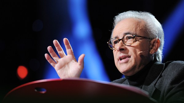
Today I want to tell you about a project being carried out by scientists all over the world to paint a neural portrait of the human mind. And the central idea of this work is that the human mind and brain is not a single, general-purpose processor, but a collection of highly specialized components, each solving a different specific problem, and yet collectively making up who we are as human beings and thinkers. To give you a feel for this idea,
오늘 저는 여러분께 전세계 과학자들이 협력하고 있는 프로젝트에 대해 말씀드리고자 합니다. 인간 사고의 신경 초상을 그리는 것입니다. 이 연구의 핵심적인 발상은 사람의 사고와 뇌가 하나의 다목적 프로세서가 아니라 아주 특화된 부분들의 집합체이며 각 부분은 서로 다른 특정 기능이 있지만 그게 다 합쳐져서 우리가 어떤 사람인지, 어떤 생각을 하는지 결정한다는 것입니다. 여러분의 이해를 위해서
imagine the following scenario: You walk into your child's day care center. As usual, there's a dozen kids there waiting to get picked up, but this time, the children's faces look weirdly similar, and you can't figure out which child is yours. Do you need new glasses? Are you losing your mind? You run through a quick mental checklist. No, you seem to be thinking clearly, and your vision is perfectly sharp. And everything looks normal except the children's faces. You can see the faces, but they don't look distinctive, and none of them looks familiar, and it's only by spotting an orange hair ribbon that you find your daughter.
다음 시나리오를 상상해보죠. 여러분이 여러분 아이의 놀이방에 들어섰습니다. 평소처럼 여러 아이가 부모를 기다리고 있는데 이번에는 아이들 얼굴이 이상하게 비슷해보입니다. 누가 여러분의 아이인지 구별을 할 수 없습니다. 새 안경이 필요한 걸까요? 정신이 이상해진 걸까요? 여러분은 머릿속으로 재빨리 확인해봅니다. 아니, 정신은 또렷한 것 같고 시야도 완벽하게 선명합니다. 모든 게 정상처럼 보여요. 아이들 얼굴만 빼고는요. 여러분은 얼굴을 볼 수는 있지만 구별할 수 없습니다. 어느 얼굴도 익숙해 보이지 않고 주황색 머리 리본을 발견하고서야 여러분의 딸을 찾습니다.
This sudden loss of the ability to recognize faces actually happens to people. It's called prosopagnosia, and it results from damage to a particular part of the brain. The striking thing about it is that only face recognition is impaired; everything else is just fine.
얼굴을 인식하는 능력을 갑자기 잃어버리는 이 현상은 실제로 일어납니다. 얼굴인식 불능증이라고 하는데 뇌의 특정한 부분이 손상된 결과입니다. 이 증상의 놀라운 점은 얼굴 인식 장애 외에는 모든 인지 기능이 정상이라는 것입니다.
Prosopagnosia is one of many surprisingly specific mental deficits that can happen after brain damage. These syndromes collectively have suggested for a long time that the mind is divvied up into distinct components, but the effort to discover those components has jumped to warp speed with the invention of brain imaging technology, especially MRI. So MRI enables you to see internal anatomy at high resolution, so I'm going to show you in a second a set of MRI cross-sectional images through a familiar object, and we're going to fly through them and you're going to try to figure out what the object is. Here we go.
얼굴인식 불능증은 뇌 손상으로 일어날 수 있는 놀랍도록 특정적인 정신 결핍 증세 중의 하나입니다. 오랜 기간 이런 증상들은 우리의 뇌 기능이 뚜렷히 구분된 요소로 이루어졌음을 시사했고 그 구성 요소들을 찾으려는 노력은 뇌 영상 기술의 발명, 특히 MRI와 함께 급속도로 발전했습니다. MRI는 신체 내부 구조를 고해상도로 보여줍니다. 잠시 후에 여러분께도 익숙할 한 물체의 MRI 단면도를 보여드리겠습니다. 연속적으로 보여드릴텐데 무엇인지 맞춰보세요. 여기 있습니다.
It's not that easy. It's an artichoke.
쉽지 않죠. 아티초크입니다.
Okay, let's try another one, starting from the bottom and going through the top. Broccoli! It's a head of broccoli. Isn't it beautiful? I love that.
좋아요. 다른 것을 보죠. 밑에서 시작해서 위로 올라갑니다. 브로콜리! 브로콜리 한덩이에요. 아름답지 않나요? 좋아요.
Okay, here's another one. It's a brain, of course. In fact, it's my brain. We're going through slices through my head like that. That's my nose over on the right, and now we're going over here, right there.
자, 또 다른 게 있어요. 당연히 뇌입니다. 사실은 제 뇌입니다. 제 뇌를 저렇게 한 단면씩 보는거죠. 오른쪽으로 제 코가 있고 지금은 이쪽으로, 바로 여기입니다.
So this picture's nice, if I do say so myself, but it shows only anatomy. The really cool advance with functional imaging happened when scientists figured out how to make pictures that show not just anatomy but activity, that is, where neurons are firing. So here's how this works. Brains are like muscles. When they get active, they need increased blood flow to supply that activity, and lucky for us, blood flow control to the brain is local, so if a bunch of neurons, say, right there get active and start firing, then blood flow increases just right there. So functional MRI picks up on that blood flow increase, producing a higher MRI response where neural activity goes up.
멋진 사진이네요. 제가 말하기는 그렇지만. 하지만 MRI 는 구조만 보여줍니다. 정말 멋진 발전은 과학자들이 뇌의 구조뿐 아니라 뇌신경 세포의 활동을 촬영하는 기능 영상을 개발함으로 이루어졌습니다. 그 원리를 설명하죠. 뇌는 근육과 같습니다. 뇌가 활동할 때 그 활동을 지원하는 혈류가 증가합니다. 다행히 뇌의 혈류 조절은 국지적이기 때문에 신경 세포 한 다발이, 예를 들면 여기에서 활동을 시작하고 신호를 발사 하면 그곳에만 혈류가 증가합니다. 기능성 MRI는 그 혈류 증가량을 감지해서 신경 활동이 증가하는 곳에서 더 높은 MRI 반응을 나타냅니다.
So to give you a concrete feel for how a functional MRI experiment goes and what you can learn from it and what you can't, let me describe one of the first studies I ever did. We wanted to know if there was a special part of the brain for recognizing faces, and there was already reason to think there might be such a thing based on this phenomenon of prosopagnosia that I described a moment ago, but nobody had ever seen that part of the brain in a normal person, so we set out to look for it. So I was the first subject. I went into the scanner, I lay on my back, I held my head as still as I could while staring at pictures of faces like these and objects like these and faces and objects for hours. So as somebody who has pretty close to the world record of total number of hours spent inside an MRI scanner, I can tell you that one of the skills that's really important for MRI research is bladder control. (Laughter)
그럼 기능성 MRI 실험이 어떻게 진행되고, 그런 실험에서 무엇을 알 수 있고 알 수 없는지 제 첫 연구 중 하나를 예로 설명하겠습니다. 우리는 얼굴 인식 기능이 뇌의 특정 부분에 존재하는지 확인하기 원했습니다. 제가 아까 설명했던 얼굴인식 불능증이란 현상을 통해 그럴 가능성을 추측할 수 있었지만 정상적인 뇌에서 이전까지 확인된 적이 없었기에 우리가 찾아나섰죠. 제가 첫번째 실험대상이었어요. 스캐너 속으로 들어가 누워서 될 수 있는대로 머리를 움직이지 않고 이런 얼굴들과 이런 사물들의 사진을 바라봤습니다. 이렇게 얼굴과 사물들을 몇 시간동안 봤죠. MRI 스캐너 안에서 보낸 총 시간수로 세계 기록에 가까운 기록을 가진 사람으로서 말씀드릴 수 있는 것은 MRI연구에서 정말 중요한 능력은 방광 조절입니다. (웃음)
When I got out of the scanner, I did a quick analysis of the data, looking for any parts of my brain that produced a higher response when I was looking at faces than when I was looking at objects, and here's what I saw. Now this image looks just awful by today's standards, but at the time I thought it was beautiful. What it shows is that region right there, that little blob, it's about the size of an olive and it's on the bottom surface of my brain about an inch straight in from right there. And what that part of my brain is doing is producing a higher MRI response, that is, higher neural activity, when I was looking at faces than when I was looking at objects. So that's pretty cool, but how do we know this isn't a fluke? Well, the easiest way is to just do the experiment again. So I got back in the scanner, I looked at more faces and I looked at more objects and I got a similar blob, and then I did it again and I did it again and again and again, and around about then I decided to believe it was for real. But still, maybe this is something weird about my brain and no one else has one of these things in there, so to find out, we scanned a bunch of other people and found that pretty much everyone has that little face-processing region in a similar neighborhood of the brain.
스캐너에서 나온 저는 간단한 데이터 분석으로 제가 사물을 볼 때보다 얼굴을 봤을 때 더 높은 반응을 일으키는 뇌 부분을 찾아보았습니다. 이것이 제가 본 거에요. 요즘 기준에서는 정말 구식 영상이지만 그때는 아름답다고 생각했어요. 이 영상에서 나타나는 바로 여기, 작은 덩어리요. 올리브 한 알의 크기인데 제 뇌의 아랫쪽에 있고 여기서 안으로 2.5 cm 들어간 곳입니다. 제 뇌의 바로 저 부분이 제가 물체를 볼 때보다 얼굴을 봤을 때 더 높은 MRI 반응, 즉, 더 높은 신경 활동을 보여주었습니다. 정말 멋진 결과였지만 우연일 수도 있지 않나요? 가장 쉬운 확인 방법은 그냥 실험을 다시 하는 거죠. 그래서 저는 스캐너 안에 들어가서 더 많은 얼굴과 더 많은 사물을 봤고 비슷한 부분이 영상에 나타났습니다. 그 뒤에 실험을 또 하고 다시 하고 다시, 또 다시 하고 그러고 나서야 결과가 진짜임을 믿기로 했죠. 하지만 어쩌면 이것은 제 뇌만 이상한 것이고 다른 사람은 이런 게 없을 지도 모릅니다. 그래서 여러 사람들을 스캔했고 거의 대부분의 사람들에서 뇌의 비슷한 부분에 얼굴을 인식하는 작은 영역이 나타났습니다.
So the next question was, what does this thing really do? Is it really specialized just for face recognition? Well, maybe not, right? Maybe it responds not only to faces but to any body part. Maybe it responds to anything human or anything alive or anything round. The only way to be really sure that that region is specialized for face recognition is to rule out all of those hypotheses. So we spent much of the next couple of years scanning subjects while they looked at lots of different kinds of images, and we showed that that part of the brain responds strongly when you look at any images that are faces of any kind, and it responds much less strongly to any image you show that isn't a face, like some of these.
그래서 다음 질문은 이 부분은 정확히 무엇을 하는걸까? 정말로 얼굴 인식에 특화된 곳인가? 아닐지도 모르겠죠? 어쩌면 얼굴뿐 아니라 아무 신체 부위나 어쩌면 사람이나 살아있는 것, 동그란 것에 반응하는지도 모릅니다. 이 부분이 얼굴 인식에 특화되었음을 확신할 유일한 방법은 그런 모든 가정을 배제하는 것이었어요. 그래서 우리는 이후 몇 년 동안 실험자들이 여러 사진을 보는 동안 뇌를 촬영했습니다. 뇌의 그 부분은 어떤 종류든 얼굴의 영상에 강하게 반응했고 이런 사진들처럼 얼굴이 아닌 것을 볼 때는 훨씬 덜 반응했습니다.
So have we finally nailed the case that this region is necessary for face recognition? No, we haven't. Brain imaging can never tell you if a region is necessary for anything. All you can do with brain imaging is watch regions turn on and off as people think different thoughts. To tell if a part of the brain is necessary for a mental function, you need to mess with it and see what happens, and normally we don't get to do that. But an amazing opportunity came about very recently when a couple of colleagues of mine tested this man who has epilepsy and who is shown here in his hospital bed where he's just had electrodes placed on the surface of his brain to identify the source of his seizures. So it turned out by total chance that two of the electrodes happened to be right on top of his face area. So with the patient's consent, the doctors asked him what happened when they electrically stimulated that part of his brain. Now, the patient doesn't know where those electrodes are, and he's never heard of the face area. So let's watch what happens. It's going to start with a control condition that will say "Sham" nearly invisibly in red in the lower left, when no current is delivered, and you'll hear the neurologist speaking to the patient first. So let's watch.
그래서 최종적으로 이 부분이 얼굴 인식에 필요하다는 결론을 내렸을까요? 아니오. 그렇지 못했습니다. 뇌 영상은 어떤 영역이 특정 용도에 필요하다는 것은 결코 알려주지 못합니다. 뇌 영상으로 할 수 있는 일은 사람들이 다른 생각을 할 때 어떤 영역들이 깜빡이는지 보는 거죠. 뇌의 한 부분이 정신 기능에 필요한지를 알려면 그 부분을 방해해 봐야 하는데 보통 그런 기회는 없습니다. 하지만 아주 최근 놀라운 기회가 생겼습니다. 제 동료 몇 명이 간질을 앓는 이 남자분을 실험했을 때였습니다. 여기 병원 침대에 누워있는 분입니다. 그의 뇌 표면에 전극을 연결해 발작의 원인을 찾으려고 했습니다. 그런데 아주 우연하게도 전극 2개가 얼굴 인식 영역 바로 위에 놓이게 되었습니다. 그래서 환자의 동의하에 의사들은 그에게 그 뇌 부분에 전기 자극이 가해졌을 때 어땠는지 물었습니다. 자, 환자는 그 전극들이 어디에 있는지 모르고 얼굴 인식 영역을 들어본 적도 없어요. 무슨 일이 일어나는지 보시죠. 비교 조건으로 시작할 텐데 전류가 흐르지 않을 때는 "가짜"라고 거의 보이지 않게 왼쪽 아래에 빨간색으로 나타날거에요. 신경학자가 환자에게 먼저 말하는 것을 들을 겁니다. 보시죠.
(Video) Neurologist: Okay, just look at my face and tell me what happens when I do this. All right?
(영상) 신경학자: 제 얼굴을 보고 이렇게 할 때 어떤지 말해주세요. 좋습니까?
Patient: Okay.
환자: 좋아요.
Neurologist: One, two, three.
신경학자: 하나, 둘, 셋.
Patient: Nothing. Neurologist: Nothing? Okay. I'm going to do it one more time. Look at my face. One, two, three.
환자: 아무일도 없어요. 신경학자: 아무일도요? 좋아요. 한 번 더 하겠습니다. 제 얼굴을 보세요. 하나, 둘, 셋.
Patient: You just turned into somebody else. Your face metamorphosed. Your nose got saggy, it went to the left. You almost looked like somebody I'd seen before, but somebody different. That was a trip. (Laughter)
환자: 의사 선생님이 다른 사람으로 바뀌었어요. 선생님 얼굴이 변했어요. 코가 축 처져서 왼쪽으로 갔어요. 제가 이전에 본 사람하고 비슷했는데 다른 사람이었어요. 마약 환각 같았어요. (웃음)
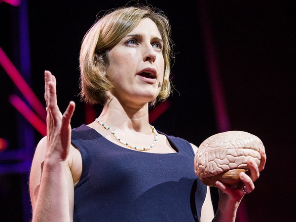
Nancy Kanwisher: So this experiment — (Applause) — this experiment finally nails the case that this region of the brain is not only selectively responsive to faces but causally involved in face perception. So I went through all of these details about the face region to show you what it takes to really establish that a part of the brain is selectively involved in a specific mental process. Next, I'll go through much more quickly some of the other specialized regions of the brain that we and others have found. So to do this, I've spent a lot of time in the scanner over the last month so I can show you these things in my brain.
낸시 캔위셔: 그래서 이 실험은 (박수) 이 실험을 통해 드디어 우리는 뇌의 이 영역이 얼굴에 선별적으로 반응할 뿐 아니라 얼굴 인식의 근원이라는 결론을 내렸습니다. 지금까지 얼굴 인식 영역을 예로 뇌의 어느 부위가 어떤 인지 능력에 선별적으로 관련이 있는지 확인하는 과정을 보여드렸습니다. 그럼 저와 다른 학자들이 찾아낸 뇌의 다른 전문 영역들에 대해 좀 더 빠르게 설명하겠습니다. 이 강연을 위해 저는 지난 달 스캐너에서 많은 시간을 보냈고 제 뇌 속 영역들을 보여드릴 수 있습니다.
So let's get started. Here's my right hemisphere. So we're oriented like that. You're looking at my head this way. Imagine taking the skull off and looking at the surface of the brain like that. Okay, now as you can see, the surface of the brain is all folded up. So that's not good. Stuff could be hidden in there. We want to see the whole thing, so let's inflate it so we can see the whole thing. Next, let's find that face area I've been talking about that responds to images like these. To see that, let's turn the brain around and look on the inside surface on the bottom, and there it is, that's my face area. Just to the right of that is another region that is shown in purple that responds when you process color information, and near those regions are other regions that are involved in perceiving places, like right now, I'm seeing this layout of space around me and these regions in green right there are really active. There's another one out on the outside surface again where there's a couple more face regions as well. Also in this vicinity is a region that's selectively involved in processing visual motion, like these moving dots here, and that's in yellow at the bottom of the brain, and near that is a region that responds when you look at images of bodies and body parts like these, and that region is shown in lime green at the bottom of the brain.
그럼 시작해볼까요. 제 뇌의 우측반구입니다. 이렇게 제 머리를 이쪽에서 보고 계십니다. 두개골을 벗겨내고 뇌 표면을 이렇게 본다고 상상해보세요. 여러분이 보다시피 뇌 표면은 모두 접혀 있습니다. 안되겠죠. 뭔가 숨어있을 수도 있으니까요. 우리는 전체를 보고 싶으니까 뇌를 부풀려서 전체를 보죠. 다음은 이런 이미지에 반응하는 얼굴 영역에 대해 찾아봅시다. 뇌를 돌려서, 아래쪽 내부 표면을 볼까요. 바로 저기가 제 얼굴 영역입니다. 거기 오른쪽에 보라색으로 보이는 다른 영역은 색깔 정보를 처리할 때 반응하고 그 근처의 다른 영역은 장소를 인식하는데 관여합니다. 지금 제가 이렇게 제 주위의 공간 배치를 보면 저기 초록색으로 보이는 영역이 왕성하게 활동합니다. 장소 영역은 뇌 표면에 하나 더 있고 거기에 얼굴 영역도 두개 더 있습니다. 또한 이 근처의 영역은 이런 움직이는 점 같은 시각적 운동을 인식하는데 선별적으로 관여하고 아래쪽 노란색으로 표시된 부분입니다. 그 근처의 영역은 몸이나 몸 부위의 이미지를 볼 때 반응합니다. 저기 뇌 아래쪽에 보시는 밝은 초록색 부분입니다.
Now all these regions I've shown you so far are involved in specific aspects of visual perception. Do we also have specialized brain regions for other senses, like hearing? Yes, we do. So if we turn the brain around a little bit, here's a region in dark blue that we reported just a couple of months ago, and this region responds strongly when you hear sounds with pitch, like these. (Sirens) (Cello music) (Doorbell) In contrast, that same region does not respond strongly when you hear perfectly familiar sounds that don't have a clear pitch, like these. (Chomping) (Drum roll) (Toilet flushing)
여태까지 제가 보여드린 영역들은 모두 시지각의 특정 측면에 관여합니다. 듣기와 같은 다른 감각에 특화된 뇌 영역도 있을까요? 예, 그렇습니다. 뇌를 약간만 돌려보면 여기 짙은 청색으로 표시된 영역을 저희 팀이 몇달전에 발표했는데 이 영역은 음정있는 소리를 들을 때 이것들처럼 강하게 반응합니다. (사이렌) (첼로 음악) (초인종) 반대로 이 영역은 아주 익숙한 소리라도 선명한 음정이 없으면 강하게 반응하지 않습니다. 예를 들면 (박수 소리) (드럼 소리) (화장실 물 내리는 소리)
Okay. Next to the pitch region is another set of regions that are selectively responsive when you hear the sounds of speech.
이 음정 영역 옆에는 말소리에 선별적으로 반응하는 영역이 있습니다.
Okay, now let's look at these same regions. In my left hemisphere, there's a similar arrangement — not identical, but similar — and most of the same regions are in here, albeit sometimes different in size.
이제 제 뇌 좌측 반구의 같은 부분을 보면 똑같지는 않지만 비슷합니다. 이 영역들은 대부분 크기만 다르게 좌우반구 둘다에 존재합니다.
Now, everything I've shown you so far are regions that are involved in different aspects of perception, vision and hearing. Do we also have specialized brain regions for really fancy, complicated mental processes? Yes, we do. So here in pink are my language regions. So it's been known for a very long time that that general vicinity of the brain is involved in processing language, but we showed very recently that these pink regions respond extremely selectively. They respond when you understand the meaning of a sentence, but not when you do other complex mental things, like mental arithmetic or holding information in memory or appreciating the complex structure in a piece of music.
제가 여태까지 보여드린 내용은 모두 감각의 관한 것이었습니다. 시각과 청각이요. 아주 고차원의 복잡한 정신 과정에 특화된 뇌의 영역도 있을까요? 예, 있습니다. 여기 분홍색은 언어 영역입니다. 오랫동안 뇌의 대부분이 언어 처리와 관련있다고 알려졌는데 아주 최근에야 이 분홍색 영역들의 매우 특정한 반응이 발견되었습니다. 이 영역들은 문장을 이해할 때는 반응하지만 다른 복잡한 사고를 할 때는 반응하지 않습니다. 암산이나, 정보를 기억하거나 음악에서 복잡한 구조를 감상할 때죠.
The most amazing region that's been found yet is this one right here in turquoise. This region responds when you think about what another person is thinking. So that may seem crazy, but actually, we humans do this all the time. You're doing this when you realize that your partner is going to be worried if you don't call home to say you're running late. I'm doing this with that region of my brain right now when I realize that you guys are probably now wondering about all that gray, uncharted territory in the brain, and what's up with that?
발견된 것 중 가장 놀라운 영역은 청록색으로 보이는 이곳입니다. 이 영역은 여러분이 다른 사람이 무엇을 생각할지 생각할 때 반응합니다. 터무니없는 것같지만 우리 인간들이 늘상 하는 일입니다. 여러분이 집에 늦게 들어갈 때 가족에게 전화하지 않으면 걱정할거라고 생각할 때, 그리고 저도 지금 그 영역을 사용하고 있습니다. 지금쯤 여러분들은 아직 밝혀지지 않은 저 회색 부분들에 대해 궁금해 할 거라고 생각하거든요. 대체 무슨 영역일까요?
Well, I'm wondering about that too, and we're running a bunch of experiments in my lab right now to try to find a number of other possible specializations in the brain for other very specific mental functions. But importantly, I don't think we have specializations in the brain for every important mental function, even mental functions that may be critical for survival. In fact, a few years ago, there was a scientist in my lab who became quite convinced that he'd found a brain region for detecting food, and it responded really strongly in the scanner when people looked at images like this. And further, he found a similar response in more or less the same location in 10 out of 12 subjects. So he was pretty stoked, and he was running around the lab telling everyone that he was going to go on "Oprah" with his big discovery. But then he devised the critical test: He showed subjects images of food like this and compared them to images with very similar color and shape, but that weren't food, like these. And his region responded the same to both sets of images. So it wasn't a food area, it was just a region that liked colors and shapes. So much for "Oprah."
저도 그게 궁금해서 제 실험실에서 일련의 실험을 통해 다른 특정 정신 기능에 특화되었을 수도 있는 뇌 부분들을 알아내려고 합니다. 하지만 저는 모든 주요 정신 기능이 뇌의 어느 특정 부분에 한정되어 있다고 생각하지는 않습니다. 생존에 필수적인 정신 기능들 조차도요. 사실 몇년전 제 실험실에 있던 한 과학자는 자신이 음식을 감지하는 뇌 영역을 발견했다고 굳게 믿은 적이 있습니다. 그 영역은 사람들이 이와 같은 이미지를 봤을 때 강하게 반응했죠. 더 나아가, 피실험자 12명중 10명이 거의 같은 곳에서 비슷한 반응을 보였습니다. 그는 한껏 고무되어서 실험실을 돌아다니며 이 커다란 발견으로 "오프라 쇼"에 나갈 거라고 얘기했어요. 하지만 후에 그는 결정적인 실험을 하게 됩니다. 이와 같은 음식 사진을 피실험자에게 보여주고 색깔과 모양은 아주 비슷하지만 음식이 아닌 사진을 보여준 뒤 비교를 했습니다. 그 영역은 두 가지 사진에 똑같이 반응했습니다. 음식 영역이 아니라 단순히 색깔과 모양을 좋아하는 영역이었어요. "오프라 쇼"는 못 나가겠죠.
But then the question, of course, is, how do we process all this other stuff that we don't have specialized brain regions for? Well, I think the answer is that in addition to these highly specialized components that I've been describing, we also have a lot of very general- purpose machinery in our heads that enables us to tackle whatever problem comes along. In fact, we've shown recently that these regions here in white respond whenever you do any difficult mental task at all — well, of the seven that we've tested. So each of the brain regions that I've described to you today is present in approximately the same location in every normal subject. I could take any of you, pop you in the scanner, and find each of those regions in your brain, and it would look a lot like my brain, although the regions would be slightly different in their exact location and in their size.
하지만 그렇다면 이제 의문점은 특화된 뇌 영역이 없는 기능들은 다 어떻게 처리되는가, 하는 것입니다. 제 생각에 그에 대한 답은 뇌에는 제가 설명한 아주 특화된 부분들 외에도 아주 다목적인 부분들이 많이 있어서 우리가 어떤 문제든 처리할 수 있다는 것입니다. 사실 우리는 최근에 여기 흰색 영역이 무엇이든 어려운 정신 노동에 반응함을 발견했습니다. 피실험자 7명한테 같은 결과가 나왔죠. 제가 오늘 설명드린 각각의 뇌영역은 모든 건강한 피실험자한테서 거의 같은 곳에 나타납니다. 여러분 중 어떤 분이라도 스캐너에 넣으면 이 모든 영역들을 찾을 수 있을겁니다. 제 뇌와 아주 비슷할 거에요. 영역들의 위치와 크기는 조금씩 다를테지만요.
What's important to me about this work is not the particular locations of these brain regions, but the simple fact that we have selective, specific components of mind and brain in the first place. I mean, it could have been otherwise. The brain could have been a single, general-purpose processor, more like a kitchen knife than a Swiss Army knife. Instead, what brain imaging has delivered is this rich and interesting picture of the human mind. So we have this picture of very general-purpose machinery in our heads in addition to this surprising array of very specialized components.
제게 이 일이 중요한 이유는 이 뇌 영역들의 위치 자체보다 애초부터 우리의 사고와 뇌가 선별적, 특정적 부분들로 이루어져 있다는 단순한 사실입니다. 안 그럴 수도 있었어요. 뇌 전체가 하나의 다용도 프로세서로서 스위스 군인칼보다는 일반적인 부엌칼과 같을 수도 있었겠죠. 그러나 뇌 영상이 보여준 것은 사람 두뇌의 다채롭고 흥미로운 모습입니다. 우리는 머릿속에 다목적 기계를 갖고 있고 그외에도 아주 특화된 일련의 구성요소들을 갖고 있어요.
It's early days in this enterprise. We've painted only the first brushstrokes in our neural portrait of the human mind. The most fundamental questions remain unanswered. So for example, what does each of these regions do exactly? Why do we need three face areas and three place areas, and what's the division of labor between them? Second, how are all these things connected in the brain? With diffusion imaging, you can trace bundles of neurons that connect to different parts of the brain, and with this method shown here, you can trace the connections of individual neurons in the brain, potentially someday giving us a wiring diagram of the entire human brain. Third, how does all of this very systematic structure get built, both over development in childhood and over the evolution of our species? To address questions like that, scientists are now scanning other species of animals, and they're also scanning human infants.
이 분야는 아직 시작 단계입니다. 인간 정신의 신경 초상화를 그리는 첫 붓질을 했을 뿐이죠. 가장 근본적인 질문들은 남아있습니다. 예를 들면 이들 각각의 영역은 정확히 무엇을 할까? 왜 세 곳의 얼굴영역과 세 곳의 장소 영역이 있고 이 부분들 사이의 일 분담은 어떠할까? 둘째, 이 모든 기능들이 뇌 안에서 어떻게 연결되었을까? 확산 영상을 사용하면 뇌 내의 여러 부분을 연결하는 신경 섬유들을 추적할 수 있습니다. 여기 보이는 방법으로 각각의 뇌 신경 세포의 연결망을 추적할 수 있습니다. 어쩌면 언젠가 인간 뇌 전체의 연결 회로도를 볼 수 있을지도요. 셋째, 이런 체계적인 구조가 어떻게 형성될까? 유아기의 성장 과정과 종의 진화 과정 둘 다에서요. 그런 질문들에 답하기 위해 현재 과학자들은 다른 종인 동물들의 뇌와 인간 유아의 뇌를 영상화 하고 있습니다.
Many people justify the high cost of neuroscience research by pointing out that it may help us someday to treat brain disorders like Alzheimer's and autism. That's a hugely important goal, and I'd be thrilled if any of my work contributed to it, but fixing things that are broken in the world is not the only thing that's worth doing. The effort to understand the human mind and brain is worthwhile even if it never led to the treatment of a single disease. What could be more thrilling than to understand the fundamental mechanisms that underlie human experience, to understand, in essence, who we are? This is, I think, the greatest scientific quest of all time.
많은 사람들이 신경과학 연구에 들어가는 많은 비용을 알츠하이머나 자폐증 같은 뇌 장애 치료에 언젠가 도움이 될 수 있다는 이유로 정당화합니다. 그것은 엄청나게 중요한 목표이고 제 연구가 어떻게든 공헌할 수 있다면 기쁘겠지만 세상에서 고장난 것을 고치는 일만 값진 일은 아닙니다. 사람의 사고와 뇌를 이해하려는 노력은 병을 하나도 못 고친다고 해도 가치가 있는 일입니다. 본질적으로 우리가 누구인지, 사람의 경험의 기저를 이루는 기본적인 구조를 이해하는 것보다 더 흥분되는 일이 어디 있겠습니까? 이것이야말로 역사상 가장 중요한 과학 탐구라고 생각합니다.
(Applause)
(박수)