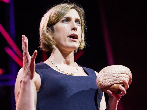
Today I want to tell you about a project being carried out by scientists all over the world to paint a neural portrait of the human mind. And the central idea of this work is that the human mind and brain is not a single, general-purpose processor, but a collection of highly specialized components, each solving a different specific problem, and yet collectively making up who we are as human beings and thinkers. To give you a feel for this idea,
היום ברצוני לספר לכם על מיזם שמבוצע בידי מדענים מכל רחבי העולם שמטרתו לצייר דיוקן עצבי של מוח האדם. והרעיון המרכזי של העבודה הזו טוען שהמוח האנושי איננו מעבד רב-תכליתי אחד, אלא אוסף של רכיבים מתמחים, כל אחד מהם פותר בעיה מסוימת, ויחד הם יוצרים את מי שאנחנו כבני אדם חושבים. כדי להמחיש לכם את הרעיון הזה,
imagine the following scenario: You walk into your child's day care center. As usual, there's a dozen kids there waiting to get picked up, but this time, the children's faces look weirdly similar, and you can't figure out which child is yours. Do you need new glasses? Are you losing your mind? You run through a quick mental checklist. No, you seem to be thinking clearly, and your vision is perfectly sharp. And everything looks normal except the children's faces. You can see the faces, but they don't look distinctive, and none of them looks familiar, and it's only by spotting an orange hair ribbon that you find your daughter.
דמיינו את התרחיש הבא: אתם נכנסים לגן הילדים של בִתכם. כרגיל, יש שם עוד עשרה ילדים שמחכים שיבואו לקחת אותם, אבל הפעם, פני הילדים נראים דומים באופן מוזר, ואתם לא מצליחים להבין מי מהם היא ילדתכם. האם אתם זקוקים למשקפיים? האם יצאתם מדעתכם? אתם בודקים את עצמכם בזריזות. לא, נראה שאתם עדיין שפויים, והראייה שלכם חדה ומדויקת. והכל נראה תקין פרט לפני הילדים. אתם רואים את הפנים, אבל אין בינם הבדלים, ואף אחד מהם לא נראה מוכר, ורק כאשר אתם מזהים את סרט השער הכתום אתם מוצאים את בתכם.
This sudden loss of the ability to recognize faces actually happens to people. It's called prosopagnosia, and it results from damage to a particular part of the brain. The striking thing about it is that only face recognition is impaired; everything else is just fine.
אובדן פתאומי של היכולת לזהות פנים קורה באמת לאנשים. המצב מכונה פרוסופגנוזיה, והוא נובע מנזק לחלק ספציפי במוח. הממצא המפתיע במצב הזה הוא שרק זיהוי הפנים נפגע, כל שאר תפקודי המוח פועלים כרגיל.
Prosopagnosia is one of many surprisingly specific mental deficits that can happen after brain damage. These syndromes collectively have suggested for a long time that the mind is divvied up into distinct components, but the effort to discover those components has jumped to warp speed with the invention of brain imaging technology, especially MRI. So MRI enables you to see internal anatomy at high resolution, so I'm going to show you in a second a set of MRI cross-sectional images through a familiar object, and we're going to fly through them and you're going to try to figure out what the object is. Here we go.
פרוסופגנוזיה הוא אחד ממספר רב של מצבי מחלה ספציפיים שיכולים להופיע אחרי פגיעה מוחית. באופן כללי מצבים אלו הצביעו על כך שהמוח מחולק לאזורים יחודיים, אבל הניסיון לזהות את אותם החלקים צבר מהירות עצומה עם המצאת טכנולוגיות להדמיית המוח, במיוחד טכנולוגיית ה- MRI (סורק תהודה מגנטית). MRI מאפשר לנו לראות את המבנה הפנימי ברזולוציה גבוהה, אראה לכם מיד סדרת תמונות חתך של MRI של חפץ מוכר ואנחנו נעבור עליהם ואתם תנסו לזהות את החפץ המדובר. בואו נתחיל
It's not that easy. It's an artichoke.
זה לא קל. מדובר בארטישוק.
Okay, let's try another one, starting from the bottom and going through the top. Broccoli! It's a head of broccoli. Isn't it beautiful? I love that.
בואו ננסה חפץ נוסף, מהבסיס ועד לקצה העליון. ברוקולי! זה ראש ברוקולי. זה לא מדהים? אני מתה על זה.
Okay, here's another one. It's a brain, of course. In fact, it's my brain. We're going through slices through my head like that. That's my nose over on the right, and now we're going over here, right there.
והנה עוד אחד. זה מוח כמובן. בעצם, זה המוח שלי. אנחנו מביטים בחתכים של הראש שלי. זה האף שלי מימין, וכאן אנחנו נמצאים כאן, בדיוק כאן.
So this picture's nice, if I do say so myself, but it shows only anatomy. The really cool advance with functional imaging happened when scientists figured out how to make pictures that show not just anatomy but activity, that is, where neurons are firing. So here's how this works. Brains are like muscles. When they get active, they need increased blood flow to supply that activity, and lucky for us, blood flow control to the brain is local, so if a bunch of neurons, say, right there get active and start firing, then blood flow increases just right there. So functional MRI picks up on that blood flow increase, producing a higher MRI response where neural activity goes up.
התמונות יפות, אם יורשה לי להחמיא לעצמי, אבל הן מראות רק את המבנה האנטומי. היתרון המדהים של הדמיה תפקודית התברר כשמדענים מצאו דרך להראות לא רק מבנה, אלא גם פעילות, כלומר, אילו תאי עצב פעילים. וככה זה עובד. המוח דומה לשרירים. כאשר התאים עובדים, הם זקוקים לזרימת דם מוגברת עבור הפעילות, ולמזלנו, השליטה בזרימת הדם למוח נעשית ברמה המקומית, כך שאם מספר תאי עצב, נגיד, אלו כאן מתחילים לפעול ולשדר, זרימת הדם תעלה בדיוק כאן. כך ש- MRI תפקודי מסוגל לקלוט את העלייה בזרימת הדם, ולהציג תגובת MRI מוגברת היכן שיש פעילות עצבית מוגברת.
So to give you a concrete feel for how a functional MRI experiment goes and what you can learn from it and what you can't, let me describe one of the first studies I ever did. We wanted to know if there was a special part of the brain for recognizing faces, and there was already reason to think there might be such a thing based on this phenomenon of prosopagnosia that I described a moment ago, but nobody had ever seen that part of the brain in a normal person, so we set out to look for it. So I was the first subject. I went into the scanner, I lay on my back, I held my head as still as I could while staring at pictures of faces like these and objects like these and faces and objects for hours. So as somebody who has pretty close to the world record of total number of hours spent inside an MRI scanner, I can tell you that one of the skills that's really important for MRI research is bladder control. (Laughter)
על מנת להמחיש לכם כיצד מתבצע ניסוי ב- MRI תפקודי, ומה ניתן ללמוד ממנו, ומה לא, אספר לכם על אחד הניסויים הראשונים שערכתי. רצינו לדעת אם יש אזור מיוחד במוח שמזהה פנים, והייתה לנו סיבה טובה לחשוב שיש אזור כזה במוח בהתבסס על תופעת הפרוסופגנוזיה שתיארתי בתחילת ההרצאה, אבל איש לא ראה מעולם את אזור המוח הזה באדם בריא, אז החלטנו לחפש אותו. אני הייתי הנבדקת הראשונה. נכנסתי לסורק, שכבתי על הגב, החזקתי את הראש יציב ככל שיכולתי בזמן שהבטתי בתמונות פנים כאלו ובחפצים כאלו ופנים וחפצים במשך שעות. וכמישהי שמחזיקה כמעט בשיא העולמי בשעות השכיבה בתוך סורק ה- MRI, אני יכולה להעיד שאחד הכישורים, החשובים למחקר ב- MRI הוא שליטה בשלפוחית השתן. (צחוק)
When I got out of the scanner, I did a quick analysis of the data, looking for any parts of my brain that produced a higher response when I was looking at faces than when I was looking at objects, and here's what I saw. Now this image looks just awful by today's standards, but at the time I thought it was beautiful. What it shows is that region right there, that little blob, it's about the size of an olive and it's on the bottom surface of my brain about an inch straight in from right there. And what that part of my brain is doing is producing a higher MRI response, that is, higher neural activity, when I was looking at faces than when I was looking at objects. So that's pretty cool, but how do we know this isn't a fluke? Well, the easiest way is to just do the experiment again. So I got back in the scanner, I looked at more faces and I looked at more objects and I got a similar blob, and then I did it again and I did it again and again and again, and around about then I decided to believe it was for real. But still, maybe this is something weird about my brain and no one else has one of these things in there, so to find out, we scanned a bunch of other people and found that pretty much everyone has that little face-processing region in a similar neighborhood of the brain.
כשיצאתי מהסורק, עברתי בזריזות על הנתונים, וחיפשתי את האזורים במוח שהגיבו חזק יותר כשהבטתי בפנים בהשוואה לחפצים, וזה מה שראיתי. תמונות הסורק האלו נראות ממש גרוע בהשוואה לתמונות שמופקות כיום, אבל בזמנו חשבתי שהן נפלאות. הן מראות את האזור שם, את הגוש הקטן הזה, בערך בגודל של זית שנמצא בחלק התחתון של המוח שלי בערך 3 ס"מ פנימה מכאן. ומה שהחלק הזה במוח שלי עושה הוא להראות תגובה חזקה יותר ב- MRI, כלומר פעילות עצבית חזקה יותר, כשהבטתי בפנים בהשוואה להסתכלות בעצמים. זה ממש מדהים, אבל איך נדע שלא מדובר בממצא מקרי? ובכן, הדרך הקלה היא לחזור על הניסוי שוב. אז נכנסתי שוב לסורק, והבטתי בעוד פנים ועוד חפצים והתוצאה הראתה כתם דומה, ואז עשיתי זאת שוב ושוב ושוב ושוב, ובערך אז התחלתי להאמין שהממצא אמיתי. ועדיין, אולי מדובר במשהו שייחודי רק למוח שלי ולאף אחד אחר אין משהו דומה במוח, אז סרקנו מוחות של אנשים נוספים ומצאנו שכמעט לכולם יש את אותו אזור שמזהה פנים במיקום דומה במוח.
So the next question was, what does this thing really do? Is it really specialized just for face recognition? Well, maybe not, right? Maybe it responds not only to faces but to any body part. Maybe it responds to anything human or anything alive or anything round. The only way to be really sure that that region is specialized for face recognition is to rule out all of those hypotheses. So we spent much of the next couple of years scanning subjects while they looked at lots of different kinds of images, and we showed that that part of the brain responds strongly when you look at any images that are faces of any kind, and it responds much less strongly to any image you show that isn't a face, like some of these.
והשאלה הבאה שעלתה הייתה, מה בדיוק עושה האזור הזה? האם הוא מתמחה רק בזיהוי פנים? אולי לא? אולי הוא מגיב לא רק לפנים אלא גם לחלקי גוף אחרים. אולי הוא מגיב לכל דבר אנושי או כל דבר חי או כל דבר עגול. הדרך היחידה לדעת בוודאות שהאזור הזה מתמחה בזיהוי פנים היא לבדוק ולשלול את כל האפשרויות האחרות. ובמהלך רוב השנתיים הבאות סרקנו מתנדבים בזמן שהם הביטו במגוון רחב של תמונות, והראינו שאותו חלק של המוח מגיב בעצמה חזקה כשמביטים בכל תמונה של פנים מכל סוג, ומגיב חלש בהרבה לתמונות שאינן פנים, כמו אלו.
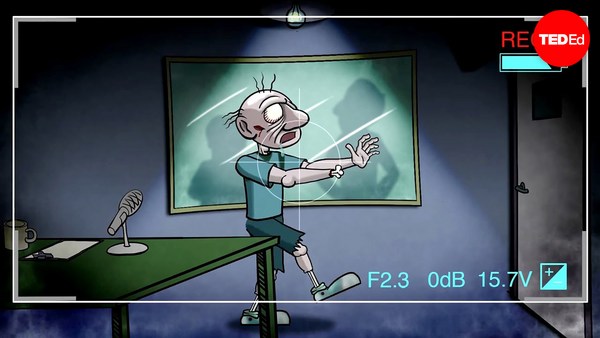
So have we finally nailed the case that this region is necessary for face recognition? No, we haven't. Brain imaging can never tell you if a region is necessary for anything. All you can do with brain imaging is watch regions turn on and off as people think different thoughts. To tell if a part of the brain is necessary for a mental function, you need to mess with it and see what happens, and normally we don't get to do that. But an amazing opportunity came about very recently when a couple of colleagues of mine tested this man who has epilepsy and who is shown here in his hospital bed where he's just had electrodes placed on the surface of his brain to identify the source of his seizures. So it turned out by total chance that two of the electrodes happened to be right on top of his face area. So with the patient's consent, the doctors asked him what happened when they electrically stimulated that part of his brain. Now, the patient doesn't know where those electrodes are, and he's never heard of the face area. So let's watch what happens. It's going to start with a control condition that will say "Sham" nearly invisibly in red in the lower left, when no current is delivered, and you'll hear the neurologist speaking to the patient first. So let's watch.
אז האם הצלחנו לקבוע בוודאות שהאזור הזה נחוץ לזיהוי פנים? לא, עדיין לא. הדמיה מוחית לא מסוגלת להוכיח אם אזור נחוץ למשהו. כל מה שהדמיית המוח עושה היא להראות אזור נדלק וכבה כשאנשים חושבים על דברים שונים. על מנת להוכיח שחלק במוח נחוץ לפעילות מוחית כלשהי, חייבים להתעסק אתו ולראות מה קורה, ובד"כ לא נותנים לנו לעשות את זה. אבל אז היה לנו מזל כאשר שני עמיתים שלי בדקו חולה אפילפסיה שמוצג כאן במיטת בית החולים מחלים מניתוח השתלת אלקטרודות על גבי המוח שלו במטרה לזהות את מקור האפילפסיה שלו. ומתברר שבמקרה 2 מהאלקטרודות מוקמו בדיוק מעל אזור זיהוי הפנים שלו. ואחרי שקיבלנו ממנו אישור הרופאים שאלו אותו מה קורה כאשר הוא מקבל גירוי חשמלי לאותו אזור במוח. חשוב להבין, החולה לא מודע למיקום האלקטרודות, והוא מעולם לא שמע על אזור זיהוי הפנים. אז בואו נראה מה קורה. הסרטון מתחיל במצב ביקורת ויהיה כתוב Sham בכתב בהיר ואדום בפינה השמאלית, כאשר לא מועבר זרם, ותשמעו את הנוירולוג מדבר עם החולה. בואו נראה.
(Video) Neurologist: Okay, just look at my face and tell me what happens when I do this. All right?
נוירולוג: תביט בפנים שלי ותגיד לי מה קורה כשאני עושה כך. בסדר?
Patient: Okay.
חולה: בסדר.
Neurologist: One, two, three.
נוירולוג: אחת, שתיים, שלוש.
Patient: Nothing. Neurologist: Nothing? Okay. I'm going to do it one more time. Look at my face. One, two, three.
חולה: כלום. נוירולוג: כלום? בסדר. בוא נעשה זאת שוב. תביט בפנים שלי. אחת, שתיים, שלוש.
Patient: You just turned into somebody else. Your face metamorphosed. Your nose got saggy, it went to the left. You almost looked like somebody I'd seen before, but somebody different. That was a trip. (Laughter)
חולה: הרגע הפכת למישהו אחר. הפנים שלך השתנו. האף שלך שקע, הוא סטה שמאלה. אתה דומה למישהו שראיתי פעם, מישהו אחר. איזה טריפ. (צחוק)
Nancy Kanwisher: So this experiment — (Applause) — this experiment finally nails the case that this region of the brain is not only selectively responsive to faces but causally involved in face perception. So I went through all of these details about the face region to show you what it takes to really establish that a part of the brain is selectively involved in a specific mental process. Next, I'll go through much more quickly some of the other specialized regions of the brain that we and others have found. So to do this, I've spent a lot of time in the scanner over the last month so I can show you these things in my brain.
ננסי קנוישר: אז הניסוי הזה - (מחיאות כפיים) - הניסוי הזה מוכיח באופן מוחלט את התאוריה שהאזור הזה במוח לא רק מגיב סלקטיבית לפנים אלא גם מראה קשר סיבתי לזיהוי הפנים. אז עברתי על כל הנתונים על אזור זיהוי הפנים שמראים את כל המידע הדרוש על מנת להוכיח שחלק זה במוח מעורב באופן ייעודי בתהליך מוחי מסוים. בהמשך, אני אעבור במהירות על האזורים המתמחים הנוספים במוח שאנחנו ואחרים גילינו. על מנת לעשות זאת, ביליתי זמן רב בתוך הסורק בחודש האחרון כדי להראות לכם את האזורים הללו במוח שלי.
So let's get started. Here's my right hemisphere. So we're oriented like that. You're looking at my head this way. Imagine taking the skull off and looking at the surface of the brain like that. Okay, now as you can see, the surface of the brain is all folded up. So that's not good. Stuff could be hidden in there. We want to see the whole thing, so let's inflate it so we can see the whole thing. Next, let's find that face area I've been talking about that responds to images like these. To see that, let's turn the brain around and look on the inside surface on the bottom, and there it is, that's my face area. Just to the right of that is another region that is shown in purple that responds when you process color information, and near those regions are other regions that are involved in perceiving places, like right now, I'm seeing this layout of space around me and these regions in green right there are really active. There's another one out on the outside surface again where there's a couple more face regions as well. Also in this vicinity is a region that's selectively involved in processing visual motion, like these moving dots here, and that's in yellow at the bottom of the brain, and near that is a region that responds when you look at images of bodies and body parts like these, and that region is shown in lime green at the bottom of the brain.
בואו נתחיל. זהו צד המוח הימני שלי. בואו נבין את הכיוונים. אתם מביטים בראשי מהכיוון הזה. דמיינו שהסרנו את הגולגולת ואנו מביטים בפני השטח של המוח כך. עכשיו תוכלו לראות, שפני השטח של המוח מקופלים. וזה לא טוב. דברים יכולים להתחבא שם. אנחנו רוצים לראות הכל, אז בואו ננפח את המוח כדי לראות את כולו. עכשיו, בואו נמצא את אזור זיהוי הפנים עליו דיברתי עד עכשיו האזור שמגיב לתמונות כאלו. כדי לראות אותו, בואו נסובב את המוח ונביט על השטח הפנימי בתחתית, והנה הוא, זהו אזור זיהוי הפנים שלי. וממש לימינו, יש אזור נוסף שמוצג בסגול שמגיב בעת עיבוד של צבעים, ולידם יש אזורים נוספים שמעורבים בתפיסת מקומות, כמו עכשיו, אני רואה את סידור הדברים במרחב סביבי והאזורים הצבועים בירוק פעילים מאד. יש עוד אזור על גבי השטח החיצוני יש שם עוד שני אזורים השותפים בזיהוי פנים. ובאותו אזור יש אזור שמעורב בעיבוד של ראיית תנועה, כמו הנקודות הנעות הללו, זה האזור הצהוב בתחתית המוח, ולידו נמצא האזור שמגיב כשמסתכלים על תמונות גוף וחלקי גוף כאלה, האזור מוצג בירוק בהיר בתחתית המוח.
Now all these regions I've shown you so far are involved in specific aspects of visual perception. Do we also have specialized brain regions for other senses, like hearing? Yes, we do. So if we turn the brain around a little bit, here's a region in dark blue that we reported just a couple of months ago, and this region responds strongly when you hear sounds with pitch, like these. (Sirens) (Cello music) (Doorbell) In contrast, that same region does not respond strongly when you hear perfectly familiar sounds that don't have a clear pitch, like these. (Chomping) (Drum roll) (Toilet flushing)
כל האזורים שהצגתי עד כה מעורבים בהיבטים שונים של תפיסת הראייה. האם יש לנו גם אזורים ספציפיים במוח לחושים האחרים, כמו שמיעה? כן, בוודאי. אם נסובב את המוח מעט, הנה אזור כחול כהה עליו דיווחנו לפני חודשיים, והאזור הזה מגיב באופן חזק כאשר שומעים קולות בגובה משתנה, כמו אלה. (סירנות) (צלילי צ'לו) (פעמון דלת) מצד שני, האזור הזה כמעט ולא מגיב כאשר אנו שומעים קולות מוכרים בלי שינוי בגובה הצליל, כאלה. (גריסה) (תופים) (שטיפת אסלה)
Okay. Next to the pitch region is another set of regions that are selectively responsive when you hear the sounds of speech.
וליד אזור גובה הצליל קיימים מספר אזורים שמגיבים באופן ייעודי לצלילים של דיבור.
Okay, now let's look at these same regions. In my left hemisphere, there's a similar arrangement — not identical, but similar — and most of the same regions are in here, albeit sometimes different in size.
בואו נביט בהם. בצד השמאלי של מוחי, קיים סידור דומה - לא זהה, אבל דומה - ורוב האזורים הדומים ממוקמים כאן, למרות הבדלים מסוימים בגודל.
Now, everything I've shown you so far are regions that are involved in different aspects of perception, vision and hearing. Do we also have specialized brain regions for really fancy, complicated mental processes? Yes, we do. So here in pink are my language regions. So it's been known for a very long time that that general vicinity of the brain is involved in processing language, but we showed very recently that these pink regions respond extremely selectively. They respond when you understand the meaning of a sentence, but not when you do other complex mental things, like mental arithmetic or holding information in memory or appreciating the complex structure in a piece of music.
כל מה שהראיתי לכם עד כה הם אזורים המעורבים בסוגים שונים של חישה: ראיה ושמיעה. האם יש גם אזורים מתמחים במוח עבור תהליכים מורכבים שמבצע המוח? כן, בודאי. כאן בורוד נמצאים אזורי השפה שלי. זו עובדה שידועה כבר הרבה שנים שהאזור הזה במוח מעורב בעיבוד השפה, אבל הראינו לאחרונה שהאזורים הורודים מגיבים בצורה מאד סלקטיבית. הם מגיבים כשאתם מבינים את משמעות המשפט, אבל לא מגיבים כשאתם עושים פעילות מוחית מורכבת אחרת, כמו חישובים או שינון מידע או מעקב אחרי המבנים המורכבים ביצירה מוזיקלית.
The most amazing region that's been found yet is this one right here in turquoise. This region responds when you think about what another person is thinking. So that may seem crazy, but actually, we humans do this all the time. You're doing this when you realize that your partner is going to be worried if you don't call home to say you're running late. I'm doing this with that region of my brain right now when I realize that you guys are probably now wondering about all that gray, uncharted territory in the brain, and what's up with that?
האזור המדהים ביותר שגיליתי עד כה הוא האזור הצבוע בטורקיז. זה אזור שמגיב כאשר אתם חושבים על מה שמישהו אחר חושב. זה נשמע מופרך, אבל למעשה, בני אדם עושים את כל הזמן. אתם עושים זאת כשאתם מבינים שבן זוגכם עלול לדאוג אם לא תתקשרו לעדכן שאתם מאחרים. אני עושה זאת עם אזור המוח שלי כרגע כאשר אני מבינה שאתם בוודאי שואלים את עצמכם עכשיו מה קורה באזור האפור, האזור הלא ממופה במוח, מה קורה שם?
Well, I'm wondering about that too, and we're running a bunch of experiments in my lab right now to try to find a number of other possible specializations in the brain for other very specific mental functions. But importantly, I don't think we have specializations in the brain for every important mental function, even mental functions that may be critical for survival. In fact, a few years ago, there was a scientist in my lab who became quite convinced that he'd found a brain region for detecting food, and it responded really strongly in the scanner when people looked at images like this. And further, he found a similar response in more or less the same location in 10 out of 12 subjects. So he was pretty stoked, and he was running around the lab telling everyone that he was going to go on "Oprah" with his big discovery. But then he devised the critical test: He showed subjects images of food like this and compared them to images with very similar color and shape, but that weren't food, like these. And his region responded the same to both sets of images. So it wasn't a food area, it was just a region that liked colors and shapes. So much for "Oprah."
גם אני שואלת את אותה שאלה, ואנחנו מנהלים כיום מספר ניסויים במעבדה שלי על מנת לנסות ולזהות מספר התמחויות אפשריות של המוח של פעילויות מוח שונות. אבל חשוב שתבינו, אני לא חושבת שיש התמחות של המוח לכל פעילות מוחית חשובה, אפילו לא לפעולות החיוניות להישרדות. למעשה, לפני מספר שנים, היה מדען במעבדה שלי שהיה משוכנע שהוא מצא את אזור המוח שמזהה מזון, והאזור הגיב בעצמה רבה בסורק כאשר נבדקים הביטו בתמונות כאלו. מעבר לכך, הוא מצא תגובה דומה באותו אזור ב-10 מתוך 12 נבדקים. כך שהוא היה משוכנע לגמרי והוא הסתובב במעבדה וסיפר לכולם שהוא יופיע בתכנית של אופרה שם יציג את התגלית שלו. אבל אז הוא תכנן את הניסוי החשוב ביותר: הוא הראה לנבדקים תמונות של מזון כמו אלה והשווה אותן לתמונות דומות מבחינת צבע וצורה, אבל לא של מזון, כמו אלו. והאזור במוח הגיב באופן דומה לשני סוגי התמונות. כך שלא מדובר באזור המזון, זה סתם אזור שאוהב צבעים וצורות. הלכה ההופעה אצל אופרה.
But then the question, of course, is, how do we process all this other stuff that we don't have specialized brain regions for? Well, I think the answer is that in addition to these highly specialized components that I've been describing, we also have a lot of very general- purpose machinery in our heads that enables us to tackle whatever problem comes along. In fact, we've shown recently that these regions here in white respond whenever you do any difficult mental task at all — well, of the seven that we've tested. So each of the brain regions that I've described to you today is present in approximately the same location in every normal subject. I could take any of you, pop you in the scanner, and find each of those regions in your brain, and it would look a lot like my brain, although the regions would be slightly different in their exact location and in their size.
אבל אז עולה השאלה, איך אנחנו מעבדים את כל סוגי המידע שאין עבורו אזור מתמחה במוח? נראה לי שהתשובה טמונה בכך שבנוסף לאזורים המתמחים אותם תיארתי, יש לנו המון אזורים במוח לעיבוד כללי, שמאפשרים לנו להתמודד עם כל בעיה בה ניתקל. למעשה, הוכחנו לאחרונה שהאזורים המסומנים כאן בלבן מגיבים בביצוע כל בעיית חשיבה קשה. לפחות מבין 7 הבעיות שבדקנו. כך שכל אזורי המוח אותם תיארתי במהלך ההרצאה היום נמצאים בערך באותם מקומות בכל נבדק נורמלי. יכולתי לקחת כל אחד מכם, להכניס אתכם לסורק, ולאתר את האזורים הללו במוח שלכם, והם ייראו מאוד בדומה למוח שלי, למרות שיהיו הבדלים קטנים במיקום המדויק ובגודל שלהם. מה שחשוב במחקר הזה
What's important to me about this work is not the particular locations of these brain regions, but the simple fact that we have selective, specific components of mind and brain in the first place. I mean, it could have been otherwise. The brain could have been a single, general-purpose processor, more like a kitchen knife than a Swiss Army knife. Instead, what brain imaging has delivered is this rich and interesting picture of the human mind. So we have this picture of very general-purpose machinery in our heads in addition to this surprising array of very specialized components.
הוא לא המיקום המדויק של אזורי המוח, אלא העובדה הפשוטה שיש לנו חלקים סלקטיביים, ספציפיים במוח. זה יכול היה להיות אחרת. המוח יכול היה לפעול כגוף אחד, מעבֵד רב-תכליתי, יותר דומה לסכין מטבח מאשר לאולר שוויצרי. אבל מה שגילינו בהדמיות המוח היא תמונה עשירה ומרתקת של המוח האנושי. כך שאנו מדמיינים את המוח כמעבד רב-תכליתי שמצוי בראשנו בתוספת למגוון מפתיע של רכיבים מתמחים.
It's early days in this enterprise. We've painted only the first brushstrokes in our neural portrait of the human mind. The most fundamental questions remain unanswered. So for example, what does each of these regions do exactly? Why do we need three face areas and three place areas, and what's the division of labor between them? Second, how are all these things connected in the brain? With diffusion imaging, you can trace bundles of neurons that connect to different parts of the brain, and with this method shown here, you can trace the connections of individual neurons in the brain, potentially someday giving us a wiring diagram of the entire human brain. Third, how does all of this very systematic structure get built, both over development in childhood and over the evolution of our species? To address questions like that, scientists are now scanning other species of animals, and they're also scanning human infants.
זוהי רק תחילת המיזם. רק החלנו לצבוע את המפה העצבית של המוח האנושי. השאלות היסודיות ביותר טרם נענו. לדוגמה, מה בדיוק עושה כל אזור כזה? מדוע דרושים לנו 3 אזורי זיהוי פנים? ושלושה אזורים למיקום, ומה חלוקת העבודה בינם? שנית, איך האזורים הללו מחוברים במוח? עם הדמיית דיפוזיה, ניתן לאתר קבוצות תאי עצב שמחברות חלקים שונים במוח, ובשיטה שרואים כאן, ניתן לאתר חיבורים של תאי עצב בודדים במוח, וייתכן שבעתיד תהיה לנו 'מפת חיווט' של כל המוח האנושי. שלישית, איך נבנית כל המערכת המסודרת הזו, גם במהלך ההתפתחות בילדות וגם במהלך התפתחות המין האנושי? על מנת לענות על שאלות כאלה, מדענים סורקים כיום מינים שונים של בעלי חיים, וגם תינוקות אנושיים.
Many people justify the high cost of neuroscience research by pointing out that it may help us someday to treat brain disorders like Alzheimer's and autism. That's a hugely important goal, and I'd be thrilled if any of my work contributed to it, but fixing things that are broken in the world is not the only thing that's worth doing. The effort to understand the human mind and brain is worthwhile even if it never led to the treatment of a single disease. What could be more thrilling than to understand the fundamental mechanisms that underlie human experience, to understand, in essence, who we are? This is, I think, the greatest scientific quest of all time.
אנשים רבים מצדיקים את ההשקעה העצומה בחקר המוח בתקווה שנגלה יום אחד כיצד לרפא מחלות מוח כמו אלצהיימר ואוטיזם. אלו מטרות חשובות להדהים, ואהיה מאושרת אם העבודה שלי תתרום למטרה זו, אבל תיקון הדברים השבורים בעולם אינו המטרה החשובה היחידה. המאמץ להבין את המוח האנושי חשוב כשלעצמו, גם אם לא נמצא טיפול לאף מחלה. מה יכול להיות מרגש יותר מאשר להבין את המנגנונים הבסיסיים המצויים בלב החוויה האנושית, להבין, בעצם, מי אנחנו? זאת, לדעתי, השאלה המדעית הגדולה ביותר של כל הזמנים.
(Applause)
(מחיאות כפיים)