Today I want to tell you about a project being carried out by scientists all over the world to paint a neural portrait of the human mind. And the central idea of this work is that the human mind and brain is not a single, general-purpose processor, but a collection of highly specialized components, each solving a different specific problem, and yet collectively making up who we are as human beings and thinkers. To give you a feel for this idea,
Je vais vous parler d’un projet qui a été mené par des scientifiques du monde entier afin de créer un portrait neuronal de l’esprit humain. Et l'idée centrale de ce travail est que l’esprit humain et le cerveau ne sont pas un processeur unique et universel, mais une collection d'éléments très spécialisés, chacun résolvant un problème spécifique et différent, mais qui collectivement constituent ce que nous sommes en tant qu’êtres humains et penseurs. Pour vous donner une idée,
imagine the following scenario: You walk into your child's day care center. As usual, there's a dozen kids there waiting to get picked up, but this time, the children's faces look weirdly similar, and you can't figure out which child is yours. Do you need new glasses? Are you losing your mind? You run through a quick mental checklist. No, you seem to be thinking clearly, and your vision is perfectly sharp. And everything looks normal except the children's faces. You can see the faces, but they don't look distinctive, and none of them looks familiar, and it's only by spotting an orange hair ribbon that you find your daughter.
imaginez le scénario suivant : vous entrez dans la garderie de votre enfant. Comme d'habitude, il y a une dizaine d'enfants attendant d’être récupérés. Mais cette fois-ci, les visages des enfants sont étrangement similaires, et vous ne savez pas lequel est le vôtre. Avez-vous besoin de lunettes ? Perdez-vous la tête ? Vous vous posez rapidement ces questions. Non, vous pensez clairement, et votre vision est parfaitement nette. Et tout semble normal, à part pour les visages de ces enfants. Vous voyez les visages, mais ils sont tous semblables, et aucun ne vous est familier, et c'est en apercevant un ruban à cheveux orange que vous trouvez votre fille.
This sudden loss of the ability to recognize faces actually happens to people. It's called prosopagnosia, and it results from damage to a particular part of the brain. The striking thing about it is that only face recognition is impaired; everything else is just fine.
Cette soudaine perte de capacité à reconnaître les visages arrive réellement aux gens. Cela s’appelle la prosopagnosie, et résulte de lésions faites à une partie bien précise du cerveau. Ce qui est intéressant est que seule la reconnaissance de visages est altérée, tout le reste fonctionne très bien.
Prosopagnosia is one of many surprisingly specific mental deficits that can happen after brain damage. These syndromes collectively have suggested for a long time that the mind is divvied up into distinct components, but the effort to discover those components has jumped to warp speed with the invention of brain imaging technology, especially MRI. So MRI enables you to see internal anatomy at high resolution, so I'm going to show you in a second a set of MRI cross-sectional images through a familiar object, and we're going to fly through them and you're going to try to figure out what the object is. Here we go.
La prosopagnosie est un des déficits mentaux étonnamment spécifiques résultant de lésions au cerveau. Ensemble, ces syndromes présupposent depuis longtemps que l'esprit est divisé en éléments distincts, et les efforts pour découvrir ces éléments ont énormément accéléré avec l'invention des technologies d'imagerie cérébrale, surtout l'IRM. L'IRM permet de voir l'anatomie interne en haute résolution, et je vais vous montrer des images en coupe transversale d'IRM d'objets communs, on va faire ça rapidement et vous allez essayer de deviner ce que c'est. Allons-y.
It's not that easy. It's an artichoke.
Ce n'est pas facile. C'est un artichaut.
Okay, let's try another one, starting from the bottom and going through the top. Broccoli! It's a head of broccoli. Isn't it beautiful? I love that.
Essayons-en une autre, allant du bas vers le haut. La tête d'un brocoli ! C'est pas beau ? J'adore ça.
Okay, here's another one. It's a brain, of course. In fact, it's my brain. We're going through slices through my head like that. That's my nose over on the right, and now we're going over here, right there.
Encore une. C'est un cerveau bien sûr. En fait, c'est mon cerveau. On traverse ma tête comme ceci. C'est mon nez à droite, et maintenant nous allons juste ici.
So this picture's nice, if I do say so myself, but it shows only anatomy. The really cool advance with functional imaging happened when scientists figured out how to make pictures that show not just anatomy but activity, that is, where neurons are firing. So here's how this works. Brains are like muscles. When they get active, they need increased blood flow to supply that activity, and lucky for us, blood flow control to the brain is local, so if a bunch of neurons, say, right there get active and start firing, then blood flow increases just right there. So functional MRI picks up on that blood flow increase, producing a higher MRI response where neural activity goes up.
C'est une belle image, je trouve, mais elle ne montre que l'anatomie. L'imagerie fonctionnelle a vraiment avancé quand les scientifiques ont créé des images montrant l'anatomie et l'activité cérébrale, montrant où les neurones sont actifs. Voici comment ça fonctionne. Le cerveau est un muscle. En s'activant, il a besoin d'un débit sanguin plus élevé, et heureusement, le débit sanguin se fait localement dans le cerveau. Donc si des neurones, disons, ici, s'activent et déchargent, le débit sanguin augmentera ici seulement. Et l'IRM fonctionnelle montrera cette augmentation du débit sanguin, montrant une réaction de l'IRM plus intense là où l'activité neuronale augmente.
So to give you a concrete feel for how a functional MRI experiment goes and what you can learn from it and what you can't, let me describe one of the first studies I ever did. We wanted to know if there was a special part of the brain for recognizing faces, and there was already reason to think there might be such a thing based on this phenomenon of prosopagnosia that I described a moment ago, but nobody had ever seen that part of the brain in a normal person, so we set out to look for it. So I was the first subject. I went into the scanner, I lay on my back, I held my head as still as I could while staring at pictures of faces like these and objects like these and faces and objects for hours. So as somebody who has pretty close to the world record of total number of hours spent inside an MRI scanner, I can tell you that one of the skills that's really important for MRI research is bladder control. (Laughter)
Pour vous montrer concrètement comment se passe une IRM fonctionnelle et ce qu'on en apprend et ce qu'on ne voit pas, je vais vous décrire une de mes premières études. Nous cherchions si une région du cerveau était associée à la reconnaissance faciale et tout nous prouvait qu'il y en avait bien une tel ce phénomène de prosopagnosie que je vous ai expliqué. Mais personne n'avait vu cette région du cerveau chez une personne normale, nous allions donc la chercher. J'ai donc été la première participante. Je me suis couchée dans le scanner, essayant de garder ma tête immobile, tout en regardant des photos de visages, comme ceux-ci, et d'objets comme ceux-ci, et des visages, et des objets, pendant des heures. Maintenant que j'ai presque battu le record du monde d'heures passées dans un scanner d'IRM, je peux vous dire qu'une compétence recherchée pour ces recherches en IRM est un parfait contrôle de sa vessie. (Rires)
When I got out of the scanner, I did a quick analysis of the data, looking for any parts of my brain that produced a higher response when I was looking at faces than when I was looking at objects, and here's what I saw. Now this image looks just awful by today's standards, but at the time I thought it was beautiful. What it shows is that region right there, that little blob, it's about the size of an olive and it's on the bottom surface of my brain about an inch straight in from right there. And what that part of my brain is doing is producing a higher MRI response, that is, higher neural activity, when I was looking at faces than when I was looking at objects. So that's pretty cool, but how do we know this isn't a fluke? Well, the easiest way is to just do the experiment again. So I got back in the scanner, I looked at more faces and I looked at more objects and I got a similar blob, and then I did it again and I did it again and again and again, and around about then I decided to believe it was for real. But still, maybe this is something weird about my brain and no one else has one of these things in there, so to find out, we scanned a bunch of other people and found that pretty much everyone has that little face-processing region in a similar neighborhood of the brain.
En sortant du scanner, j'ai fait une rapide analyse des données, cherchant une région du cerveau produisant plus de réactions en regardant des visages qu'en regardant des objets, et voici ce que j'ai vu. Pour les normes actuelles, cette image est horrible, mais à l'époque je la trouvais magnifique. Elle montre cette région juste là, cette petite goutte, de la taille d'une olive sur la surface inférieure de mon cerveau à plus ou moins 2 ou 3 cm à l'intérieur à partir d'ici. C'est cette partie de mon cerveau qui produit une plus grande réaction à l'IRM, donc plus d'activité neuronale, quand je regardais des visages que quand je regardais des objets. C'est super, mais comment être sûr qu'il n'y a pas d'erreur ? Le moyen le plus simple est de refaire l'expérience. Je suis retournée dans le scanner, j'ai regardé d'autres visages et d'autres objets et j'ai eu une même goutte, et je l'ai refait et je l'ai encore refait et encore et encore, et à ce moment-là, j'ai décidé que ça ne devait pas être une erreur. Mais c'était peut-être spécifique à mon cerveau et personne d'autre n'a cette petite goutte-là. Nous avons donc scanné d'autres personnes et vu que pratiquement tout le monde a cette région de reconnaissance faciale presqu'au même endroit.
So the next question was, what does this thing really do? Is it really specialized just for face recognition? Well, maybe not, right? Maybe it responds not only to faces but to any body part. Maybe it responds to anything human or anything alive or anything round. The only way to be really sure that that region is specialized for face recognition is to rule out all of those hypotheses. So we spent much of the next couple of years scanning subjects while they looked at lots of different kinds of images, and we showed that that part of the brain responds strongly when you look at any images that are faces of any kind, and it responds much less strongly to any image you show that isn't a face, like some of these.
La question suivante a dont été : à quoi sert cette chose ? Simplement à reconnaître des visages ? Peut-être pas ? Peut-être reconnait-il les visages mais aussi n'importe quelle partie du corps. Peut-être réagit-il à ce qui est humain ou ce qui est vivant ou ce qui est rond. Pour être sûr que cette région soit spécialisée dans la reconnaissance faciale est d'exclure ces autres hypothèses. Nous avons donc passé deux ans à scanner des gens regardant différentes images, démontrant que cette partie du cerveau réagit fortement aux images de tous types de visages, et réagit beaucoup moins aux images qui ne sont pas des visages, comme celles-ci.
So have we finally nailed the case that this region is necessary for face recognition? No, we haven't. Brain imaging can never tell you if a region is necessary for anything. All you can do with brain imaging is watch regions turn on and off as people think different thoughts. To tell if a part of the brain is necessary for a mental function, you need to mess with it and see what happens, and normally we don't get to do that. But an amazing opportunity came about very recently when a couple of colleagues of mine tested this man who has epilepsy and who is shown here in his hospital bed where he's just had electrodes placed on the surface of his brain to identify the source of his seizures. So it turned out by total chance that two of the electrodes happened to be right on top of his face area. So with the patient's consent, the doctors asked him what happened when they electrically stimulated that part of his brain. Now, the patient doesn't know where those electrodes are, and he's never heard of the face area. So let's watch what happens. It's going to start with a control condition that will say "Sham" nearly invisibly in red in the lower left, when no current is delivered, and you'll hear the neurologist speaking to the patient first. So let's watch.
Pouvons-nous maintenant être surs que cette région est nécessaire pour la reconnaissance faciale ? Non, bien sûr. L'imagerie cérébrale ne peut jamais dire si une région est nécessaire pour quoi que ce soit. L'imagerie cérébrale montre simplement si une région s'active ou pas lorsqu'on pense à différentes choses. Pour savoir si une partie du cerveau est nécessaire à une fonction mentale, vous devez la perturber et voir ce qu'il se passe, et normalement, nous ne pouvons pas faire ça. Une opportunité s'est présentée récemment lorsque deux de mes collègues ont testé cet homme épileptique, on le voit ici dans son lit d'hôpital avec des électrodes sur la surface de son cerveau pour identifier la source de ses crises. Et nous avons vraiment eu la chance que deux des électrodes soient placées exactement sur la région des visages. Avec le consentement du patient, le médecin lui demanda ce qu'il se passait quand ils stimulaient électriquement cette partie de son cerveau. Le patient ne sait bien sûr pas où sont mises les électrodes, il ne connaît pas cette « région des visages ». Regardons ce qu'il se passe. Cela commence avec un test de contrôle, le mot « Sham » [Faux] va apparaître très brièvement en rouge en bas à gauche, sans qu'aucun courant ne soit envoyé, et on entendra le neurologue parler au patient. Regardons.
(Video) Neurologist: Okay, just look at my face and tell me what happens when I do this. All right?
(Vidéo) Neurologue : Regardez mon visage et dites-moi ce qu'il se passe quand je fais ceci. D'accord ?
Patient: Okay.
Patient : Ok.
Neurologist: One, two, three.
Neurologue : Un, deux, trois.
Patient: Nothing. Neurologist: Nothing? Okay. I'm going to do it one more time. Look at my face. One, two, three.
Patient : Rien. Neurologue : Rien ? Ok. Je vais le refaire. Regardez mon visage. Un, deux, trois.
Patient: You just turned into somebody else. Your face metamorphosed. Your nose got saggy, it went to the left. You almost looked like somebody I'd seen before, but somebody different. That was a trip. (Laughter)
Patient : Vous êtes devenu quelqu'un d'autre. Votre visage s'est métamorphosé. Votre nez s'est ramolli et est allé sur la gauche. Vous ressembliez à quelqu'un que je connais, mais quelqu'un d'autre. C'était hallucinant. (Rires)
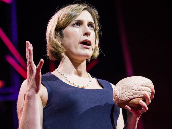
Nancy Kanwisher: So this experiment — (Applause) — this experiment finally nails the case that this region of the brain is not only selectively responsive to faces but causally involved in face perception. So I went through all of these details about the face region to show you what it takes to really establish that a part of the brain is selectively involved in a specific mental process. Next, I'll go through much more quickly some of the other specialized regions of the brain that we and others have found. So to do this, I've spent a lot of time in the scanner over the last month so I can show you these things in my brain.
Nancy : Cette expérience - (Applaudissements) - Cette expérience prouve finalement que cette région du cerveau n'est pas seulement stimulée par des visages mais sert aussi à percevoir ces visages. Je vous ai donc parlé de tout ceci sur cette région des visages pour vous montrer qu'il est difficile de prouver qu'une région du cerveau est impliquée dans un processus mental spécifique. Je vais rapidement parler maintenant d'autres régions spécialisées du cerveau que nous et d'autres avons trouvées. Pour cela, j'ai passé beaucoup de temps dans le scanner dernièrement pour vous les montrer sur mon cerveau.
So let's get started. Here's my right hemisphere. So we're oriented like that. You're looking at my head this way. Imagine taking the skull off and looking at the surface of the brain like that. Okay, now as you can see, the surface of the brain is all folded up. So that's not good. Stuff could be hidden in there. We want to see the whole thing, so let's inflate it so we can see the whole thing. Next, let's find that face area I've been talking about that responds to images like these. To see that, let's turn the brain around and look on the inside surface on the bottom, and there it is, that's my face area. Just to the right of that is another region that is shown in purple that responds when you process color information, and near those regions are other regions that are involved in perceiving places, like right now, I'm seeing this layout of space around me and these regions in green right there are really active. There's another one out on the outside surface again where there's a couple more face regions as well. Also in this vicinity is a region that's selectively involved in processing visual motion, like these moving dots here, and that's in yellow at the bottom of the brain, and near that is a region that responds when you look at images of bodies and body parts like these, and that region is shown in lime green at the bottom of the brain.
Commençons. Voici mon hémisphère droit. On est orienté comme ceci. Vous voyez ma tête comme ceci. Imaginez-la sans crâne, vous regardez la surface du cerveau comme ceci. Ok, comme vous voyez, la surface est chiffonnée. Quelque chose peut se cacher dedans. Nous voulons tout voir. Nous allons la gonfler pour la voir entièrement. Ensuite, trouvons cette région des visages qui réagit à des images comme celles-ci. Tournons donc le cerveau et regardons dans la surface inférieure, la voici, ma région des visages. À sa droite, se trouve une autre région, ici en mauve, qui réagit quand vous traitez des informations de couleurs, et près de ces régions, s'en trouvent d'autres impliquées dans la perception de lieux, comme en ce moment, je vois l'espace autour de moi et ces régions en vert ici sont très actives. Il y en a une autre à l'extérieur de cette surface où se trouvent d'autres régions des visages. Dans leur voisinage, se trouve une région impliquée dans le traitement des mouvements perçus, comme ces points mobiles ici, elle est en jaune dans le bas du cerveau, et tout près, il y a une région qui réagit quand vous regardez des images de corps, de parties de corps, comme celles-ci, elle est en vert citron sur le bas du cerveau.
Now all these regions I've shown you so far are involved in specific aspects of visual perception. Do we also have specialized brain regions for other senses, like hearing? Yes, we do. So if we turn the brain around a little bit, here's a region in dark blue that we reported just a couple of months ago, and this region responds strongly when you hear sounds with pitch, like these. (Sirens) (Cello music) (Doorbell) In contrast, that same region does not respond strongly when you hear perfectly familiar sounds that don't have a clear pitch, like these. (Chomping) (Drum roll) (Toilet flushing)
Toutes ces régions sont impliquées dans des aspects particuliers de la perception visuelle. Avons-nous aussi des régions spécialisées du cerveau pour les autres sens, comme l'ouïe ? Oui. Si nous tournons un peu le cerveau, cette région en bleu foncé, que nous avons signalée il y a quelques mois, réagit très fortement aux sons comme ceux-ci. (Sirènes) (Violoncelle) (Sonnette) Par contre, elle ne réagit pas à des sons plus familiers qui n'ont pas une sonorité claire, comme ceux-ci. (Grignotage) (Roulement de tambour) (Chasse d'eau)
Okay. Next to the pitch region is another set of regions that are selectively responsive when you hear the sounds of speech.
Ok. À côté de cette région de la sonorité, se trouvent quelques régions qui réagissent plutôt quand vous entendez des paroles.
Okay, now let's look at these same regions. In my left hemisphere, there's a similar arrangement — not identical, but similar — and most of the same regions are in here, albeit sometimes different in size.
Regardons ces régions. C'est très similaire dans mon hémisphère gauche - pas identique, mais similaire - et la plupart de ces régions s'y trouvent, parfois de tailles différentes.
Now, everything I've shown you so far are regions that are involved in different aspects of perception, vision and hearing. Do we also have specialized brain regions for really fancy, complicated mental processes? Yes, we do. So here in pink are my language regions. So it's been known for a very long time that that general vicinity of the brain is involved in processing language, but we showed very recently that these pink regions respond extremely selectively. They respond when you understand the meaning of a sentence, but not when you do other complex mental things, like mental arithmetic or holding information in memory or appreciating the complex structure in a piece of music.
Toutes ces régions sont impliquées dans différents aspects de la perception, la vision et l'ouïe. Avons-nous des régions spécialisées du cerveau pour des processus mentaux plus complexes ? Oui, bien sûr. Ici, en rose sont mes régions du langage. On sait depuis longtemps que les alentours du cerveau sont impliqués dans le traitement du langage, mais nous avons démontré récemment que ces régions en rose réagissent de manière très sélective. Elles réagissent quand vous comprenez le sens d'une phrase, mais pas en faisant d'autres choses mentalement complexes, comme du calcul mental, ou retenir des informations en mémoire ou apprécier la structure complexe d'un morceau de musique.
The most amazing region that's been found yet is this one right here in turquoise. This region responds when you think about what another person is thinking. So that may seem crazy, but actually, we humans do this all the time. You're doing this when you realize that your partner is going to be worried if you don't call home to say you're running late. I'm doing this with that region of my brain right now when I realize that you guys are probably now wondering about all that gray, uncharted territory in the brain, and what's up with that?
La région la plus surprenante trouvée pour l'instant est celle-ci en turquoise. Cette région réagit quand vous pensez à ce que pense quelqu'un d'autre. Ça peut sembler bizarre, mais en fait, nous faisons ça tout le temps. Vous le faites en vous rendant compte que votre partenaire va s'inquiéter si vous ne l'appelez pas pour prévenir d'un retard. Je le fais en ce moment même avec cette région-là en me rendant compte que vous tous, vous vous posez des questions sur tous ces territoires inconnus du cerveau et pourquoi c'est comme ça ?
Well, I'm wondering about that too, and we're running a bunch of experiments in my lab right now to try to find a number of other possible specializations in the brain for other very specific mental functions. But importantly, I don't think we have specializations in the brain for every important mental function, even mental functions that may be critical for survival. In fact, a few years ago, there was a scientist in my lab who became quite convinced that he'd found a brain region for detecting food, and it responded really strongly in the scanner when people looked at images like this. And further, he found a similar response in more or less the same location in 10 out of 12 subjects. So he was pretty stoked, and he was running around the lab telling everyone that he was going to go on "Oprah" with his big discovery. But then he devised the critical test: He showed subjects images of food like this and compared them to images with very similar color and shape, but that weren't food, like these. And his region responded the same to both sets of images. So it wasn't a food area, it was just a region that liked colors and shapes. So much for "Oprah."
Moi aussi, je me pose la question, et nous faisons d'autres tests dans mon laboratoire pour essayer d'identifier d'autres régions du cerveau spécialisées pour d'autres fonctions mentales particulières. En revanche, je pense que nous n'avons pas de zones spécialisées pour chaque fonction mentale importante, même pour les fonctions mentales importantes pour la survie. Il y a quelques années dans mon labo, ce scientifique était convaincu d'avoir trouvé une région du cerveau pour trouver de la nourriture, elle réagissait fortement au scanner quand les gens voyaient des images comme celles-ci. Il a ainsi trouvé une réaction similaire plus ou moins dans la même région chez 10 des 12 participants. Il était donc extrêmement content, courant partout, criant à tous qu'il serait chez « Oprah » pour parler de sa grande découverte. Mais, il a après conçu le test critique : il a montré aux participants des images de nourriture comme celles-ci et les a comparées à des images de couleurs et de formes similaires d'autres choses, comme celles-ci. Et sa région a réagi de la même manière aux différentes images. Ce n'était pas une région de nourriture mais une région de couleurs et de formes. Tant pis pour « Oprah. »
But then the question, of course, is, how do we process all this other stuff that we don't have specialized brain regions for? Well, I think the answer is that in addition to these highly specialized components that I've been describing, we also have a lot of very general- purpose machinery in our heads that enables us to tackle whatever problem comes along. In fact, we've shown recently that these regions here in white respond whenever you do any difficult mental task at all — well, of the seven that we've tested. So each of the brain regions that I've described to you today is present in approximately the same location in every normal subject. I could take any of you, pop you in the scanner, and find each of those regions in your brain, and it would look a lot like my brain, although the regions would be slightly different in their exact location and in their size.
La question maintenant est donc : comment traitons-nous ces autres choses pour lesquelles il n'y a pas de régions spécialisées ? Je pense qu'en plus de ces éléments très spécialisés que j'ai décrits, nous avons aussi dans nos têtes une machinerie à usage plus général qui nous permet d'affronter tous les problèmes que nous croisons. Nous avons récemment démontré que ces régions ici en blanc réagissent quand nous effectuons une tâche mentale complexe, à chaque fois - en tout cas, des 7 que nous avons testées. Donc, toutes ces régions dont j'ai parlé aujourd'hui sont présentes pratiquement au même endroit chez tous les participants normaux. Je pourrais tester quelqu'un ici, le scanner, et trouver toutes ces régions dans son cerveau, et ça ressemblerait à mon cerveau, même si ces régions se différencieraient sur leur localisation et leur taille exacte.
What's important to me about this work is not the particular locations of these brain regions, but the simple fact that we have selective, specific components of mind and brain in the first place. I mean, it could have been otherwise. The brain could have been a single, general-purpose processor, more like a kitchen knife than a Swiss Army knife. Instead, what brain imaging has delivered is this rich and interesting picture of the human mind. So we have this picture of very general-purpose machinery in our heads in addition to this surprising array of very specialized components.
Ce que je trouve très important ici n'est pas vraiment la localisation exacte de ces régions, mais le fait très simple que nous avons des régions sélectives et spécifiques dans l'esprit et le cerveau. Ça aurait pu être différent. Notre cerveau aurait pu être un seul processeur à usage général, plus comme un couteau de cuisine qu'un couteau suisse. Ce que l'imagerie cérébrale a révélé est cette image riche et intéressante de l'esprit humain. Nous avons une image de cette machinerie à usage général en plus de cette panoplie surprenante d'éléments très spécialisés.
It's early days in this enterprise. We've painted only the first brushstrokes in our neural portrait of the human mind. The most fundamental questions remain unanswered. So for example, what does each of these regions do exactly? Why do we need three face areas and three place areas, and what's the division of labor between them? Second, how are all these things connected in the brain? With diffusion imaging, you can trace bundles of neurons that connect to different parts of the brain, and with this method shown here, you can trace the connections of individual neurons in the brain, potentially someday giving us a wiring diagram of the entire human brain. Third, how does all of this very systematic structure get built, both over development in childhood and over the evolution of our species? To address questions like that, scientists are now scanning other species of animals, and they're also scanning human infants.
Nous n'en sommes encore qu'au début. Nous avons seulement apposé les premiers coups de pinceau de ce portrait neuronal de l'esprit humain. Les plus grandes questions restent sans réponse. Par exemple, quelle est la fonction exacte de ces régions ? Pourquoi avons-nous besoin de trois régions pour les visages et les lieux ? Et quelle est la division du travail entre elles ? Deuxièmement, comment toutes ces choses sont-elles connectées dans le cerveau ? Avec l'imagerie de diffusion, vous pouvez suivre des neurones qui relient entre elles différentes régions du cerveau, et avec cette méthode ici, vous pouvez suivre ces connections entre des neurones individuels, pour avoir peut-être un jour un schéma exact des connexions de tout le cerveau. Troisièmement, comment cette structure systématique se construit-elle, à travers le développement dans l'enfance et à travers l'évolution de notre espèce ? Pour répondre à ces questions, les scientifiques scannent maintenant d'autres espèces animales, ainsi que des bébés.
Many people justify the high cost of neuroscience research by pointing out that it may help us someday to treat brain disorders like Alzheimer's and autism. That's a hugely important goal, and I'd be thrilled if any of my work contributed to it, but fixing things that are broken in the world is not the only thing that's worth doing. The effort to understand the human mind and brain is worthwhile even if it never led to the treatment of a single disease. What could be more thrilling than to understand the fundamental mechanisms that underlie human experience, to understand, in essence, who we are? This is, I think, the greatest scientific quest of all time.
Beaucoup de gens justifient le coût de la recherche neuroscientifique en disant que ça nous aidera un jour à guérir des maladies comme Alzheimer et l'autisme. C'est un objectif très important, et je serais enchantée si mon travail y contribue, mais réparer ce qui est cassé dans le monde n'est pas la seule chose importante à faire. Les efforts pour comprendre l'esprit et le cerveau humain en valent la peine même s'ils n'aident jamais à guérir la moindre maladie. Quoi de plus excitant que de comprendre les mécanismes fondamentaux qui sous-tendent l'expérience humaine, de comprendre, en fait, qui nous sommes ? C'est pour moi la plus grande quête scientifique de tous les temps.
(Applause)
(Applaudissements)