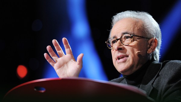
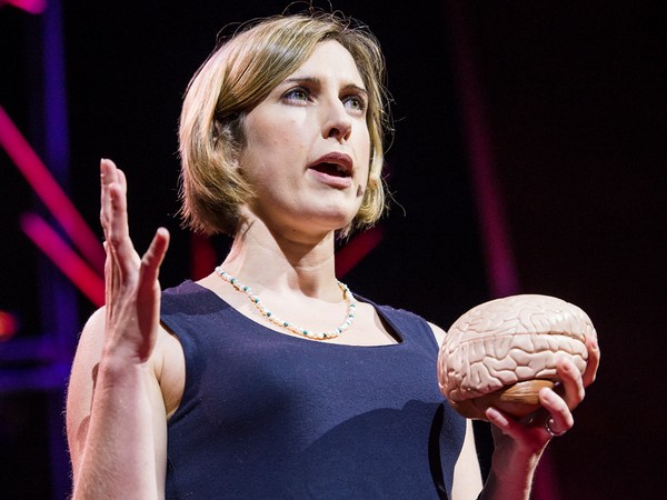
أرغب اليوم في أن أخبركم عن مشروع يشارك فيه العديد من العلماء من مختلف أنحاء العالم لرسم صورة عصبية للعقل البشريّ. الفكرة المركزية وراء هذا المشروع هي أنّ العقل والدماغ البشريّ هو ليس معالج معلومات أحاديّا عامّا بل هو مجموعة من المكونات المحدّدة بدقّة كل منها له دوره الخاص لكنها تشكّل معا في نفس الوقت هويتّنا كبشر مفكّرين. لإعطائك نبذة عن تلك الفكرة، تخيّل السيناريو التّالي: دخلت لتوّك حضانة الأطفال حيث يوجد أحد أطفالك وكالعادة، هناك عشرات من الأطفال هناك بانتظار آبائهم لكنك هذه المرّة تحسّ وكأن وجوه جميع الأطفال مألوفة ولا تستطيع تمييز أيّهم هو طفلك. هل تحتاج نظارات جديدة؟ هل أنت بصدد فقدان عقلك؟ تحاول تفقّد قدراتك الذهنية، لا، أنت تفكّر جيدا وبطريقة عادية ونظرك لا يزال حادّا كالعادة وكلّ شيء يبدو عاديّا باستثناء وجوه الأطفال. تستطيع رؤية الوجوه لكنّها لا تبدو مميّزة ولا أحد منها يبدو مألوفا ولولا رؤية أشرطة الشعر البرتقالية لما تعرّفت على ابنتك. هذا الفقدان المفاجئ لقدرة تمييز الوجوه يحدث فعلا للأشخاص. ويسمّى "عمى التعرّف على الوجوه" وهو نتيجة تلف لمنطقة محدّدة من الدماغ. الأمر المذهل هو أنّ التعرّف على الوجوه فقط يتعطّل بينما بقية الوظائف بخير. عمى التعرّف على الوجوه هو من أكثر الإعاقات الذهنية غرابة التي يمكن أن تحدث نتيجة تضرّر الدماغ. هذه المتلازمات مجتمعة كانت تشير منذ وقت طويل إلى كون العقل منقسما إلى عناصر منفصلة، لكن محاولات اكتشاف هذه العناصر تزايدت بشكل ملحوظ منذ اختراع تكنولوجيا التصوير الدماغي وخاصة التصوير بالرنين المغناطيسي. إذ أنّه يمكّن من رؤية التركيبة الداخلية بدرجة عالية من الوضوح. سوف أريكم بعد قليل مجموعة من صور مقطعية بالرنين المغناطيسي لأمر مألوف وسوف نسافر عبر الصور محاولين معرفة ما هو هذا الشيء. لنبدأ. ليس سهلا، إنّه خرشوف. حسنا، لنجرّب مرة أخرى، بدء من الأسفل وصعودا إلى أعلى القرنبيط! رأس القرنبيط أليس جميلا؟ أحبه هذا آخر، إنّه دماغ بالطبع دماغي في الواقع سوف نمرّ خلال رأسي عبر شرائح هكذا هذا أنفي إلى اليمين، والآن سوف نذهب هنا، هناك تماما الصورة جميلة، وهو أيضا رأيي الشخصي لكنها ترينا التشريح فقط. التطور المثير في التصوير الوظيفيّ حدث عندما اكتشف العلماء كيفية صنع صور لا تكشف التشريح فقط بل النشاط أيضا أي عندما تعمل الخلايا العصبية. إليكم كيف يعمل الأمر الأدمغة هي أشبه بعضلات عندما تصبح نشطة، تحتاج إلى ازدياد تدفّق الدم للتزود من أجل ذلك النشاط ومن حسن حظنا، التحكم بتدفق الدم في الدم موجود قريبا إن رغبت مجموعة من الخلايا العصبية في العمل والإرسال، يزداد تدفق الدم هناك بالذات يلتقط التصوير بالرنين المغناطيسي الوظيفي ذلك الارتفاع في ضغط الدم منتجا استجابة رنين مغناطيسي أفضل عند ارتفاع النشاط العصبي. لإعطائكم فكرة واقعية عن كيفية حدوث تجربة التصوير بالرنين المغناطيسي الوظيفي وما نتعلمه عنها أو لا نتعلمه عنها دعوني أصف إحدى أولى دراسات على الإطلاق أردنا معرفة إن كانت هناك منطقة من الدماغ وظيفتها تمييز الوجوه وكان هناك ما يدعونا للاعتقاد بذلك اعتمادا على ظاهرة عمى تعرّف الوجوه التي وصفتها منذ قليل لكن ما من أحد أجد ذلك الجزء من الدماغ في شخص عادي لذلك قررنا البحث عنه. كنت أنا أولى المجربين ذهبت للماسح، استلقيت على ظهري، أبقيت رأسي ثابتا لأطول مدة ممكنة أثناء مشاهدة صور لوجوه هكذا وأشياء كهذه وجوها وأشياء لساعات. كشخص يقترب من تحطيم الرقم القياسي لعدد الساعات المقضية داخل ماسح التصوير بالرنين المغناطيسي، أخبركم أن إحدى المهارات الهامة جدا أثناء القيام بذلك هي حبس البول. (ضحك) عند خروجي من الماسح قمت بتحليل سريع للبيانات بحثا عن أيّ أجزاء من دماغي ولّدت استجابة أقوى عند رؤيتي للوجوه مقارنة بعند رؤيتي للأشياء وهذا ما رأيته. هذه الصورة تبدو سيئة مقارنة بالمعايير الحالية لكنها بدت لي جميلة في ذلك الوقت. وهي ترينا أنّ المنطقة هنا تماما تلك النقطة الصغيرة في حجم حبة زيتون تقريبا وهي في أسفل سطح دماغي بعيدة إنشا واحدا تقريبا عن هنا ما تقوم به تلك المنطقة من الدماغ يولّد استجابة رنين مغناطيسي أقوى يعني، هناك نشاط عصبي أقوى عند رؤيتي للوجوه مقارنة بوقت رؤيتي للأجسام. الأمر رائع لكن كيف نعرف أنّها ليست ضربة حظ؟ الطريقة الأسهل هي بالقيام بالتجربة مجددا. إذن عدت للماسح ونظرت لوجوه أكثر وأشياء أكثر وحصلت على نفس النتيجة ثم كررت الأمر وأعدتها مجددا ومجددا ومجددا عندها فقط بدأت أصدّق أنّ الأمر صحيح. لكن ربما هو أمر مرتبط بدماغي أنا فقط وما من أحد له نفس الأمر وللتأكد، قمنا بمسح أدمغة أشخاص آخرين واكتشفنا أنّ جميعهم يمتلكون تلك المنطقة الخاصة بتمييز الوجوه في مكان مماثل بأدمغتهم. السؤال الذي طرح بعد ذلك هو ماهي فعلا وظيفة ذلك الشيء؟ هل هو فعلا مخصّص فقط لتمييز الوجوه؟ ربّما لا، ربما لا يستجيب للوجوه فقط بل لبقية أعضاء الجسم أيضا ربما يستجيب لأي شيء بشريّ أو أي كائن حيّ أو أي شيء دائري. الطريقة الوحيدة للتأكد أنّ تلك المنطقة مسؤولة فقط عن تمييز الوجوه هي عبر دحض تلك الافتراضات. لذلك قضينا الكثير من وقتنا في السنوات اللاحقة نمسح أدمغة أشخاص أثناء نظرهم لمختلف أنواع الصور وكشفنا أنّ ذلك الجزء من الدماغ يستجيب بقوة عند نظرك إلى أي صورة لأيّ نوع من أنواع الوجوه ويستجيب بصفة أقل بكثير لأي صورة ليست لوجه مثل هذه. إذن، هل تأكدنا نهائيا من كون تلك المنطقة ضرورية لتمييز الوجوه؟ لا، ليس بعد. التصوير الدماغي غير قادر على إخبارنا إن كانت منطقة ما ضرورية لوظيفة ما. ما يمكنك فعله بالتصوير الدماغي هو مشاهدة المناطق تعمل وتتوقف عن العمل أثناء تفكير الأشخاص بمختلف الأمور. لمعرفة ما إن كانت منطقة ما ضرورية لوظيفة ذهنية، تحتاج للعبث بها وترقّب النتيجة لكننا لا نستطيع القيام بذلك عادة. لكن فرصة مميزة ظهرت مؤخّرا عندما قام بعض زملائي باختبار شخص مصاب بداء الصرع ترونه هنا مستلقيا بالمستشفى حيث تمّ وضع أقطاب كهربائية على سطح دماغه لمعرفة مصدر نوبات صرعه. وبالصدفة، تبيّن أنّ القطبين الكهربائيين كانا موجودين في منطقة فوق رأسه تماما. إذن وبموافقة المريض، سأله الأطباء عمّا حدث عندما قاموا بتحفيز تلك المنطقة من الدماغ. المريض لم يكن يعلم مكان الأقطاب الكهربائية ولم يسبق له أن سمع بمنطقة الوجه بالدماغ. لنشاهد ما حدث. سيبدأ الأمر بظروف تحت السيطرة حيث تظهر كلمة "شام" شبه متخفية بالأحمر بأسفل الشمال حيث لا يوجد تيار كهربائي وستسمعون طبيب الأعصاب يتحدث مع المصاب أولا. لنرى. (فيديو) طبيب الأعصاب: حسنا، انظر إلى وجهي وأخبرني عمّا يحدث عندما أفعل هذا حسنا؟ المصاب: حسنا. الطبيب: واحد، اثنان، ثلاثة المصاب: لا شيء الطبيب: لا شيء؟ حسنا. سأقوم مجددا بالأمر انظر إلى وجهي واحد، اثنان، ثلاثة المصاب: لقد تحوّلت إلى شخص آخر لقد تغيّر وجهك أنفك أصبح أكبر، ومتجّها نحو الأيسر تبدو تقريبا كشخص مألوف لي لكنك شخص مختلف. يا لها من رحلة. (ضحك) نانسي كانويشر: هذه التجربة — (تصفيق) هذه التجربة تؤكد أخيرا الاعتقاد بكون هذه المنطقة من الدماغ ليست فقط تستجيب للوجوه بطريقة مختارة لكنها تتدخّل في رؤية الوجوه. لقد مررت بكل هذه التفاصيل لأكشف لكم ما يتطلّبه الأمر قبل الإقرار بأنّ منطقة من الدماغ هي بالفعل مسؤولة عن وظيفة ذهنية معيّنة. الآن سوف أمرّ بطريقة أسرع ببعض المناطق الأخرى من الدماغ التي اكتشفتها أنا وغيري. للقيام بذلك، قضيت الكثير من الوقت في الماسح أثناء الشهر المنصرم حتّى أريكم هذه الأشياء بدماغي. لنبدأ. هذا نصف الكرة المخية اليمنى الخاص بي نحن ننظر بهذا الاتجاه. أنتم تنظرون لرأسي هكذا. تخيّل نزع الجمجمة والنظر إلى سطح الدماغ هكذا. الآن يمكنكم رؤية سطح الدماغ مطويّ، وهذا سيء يمكن لأشياء أن تكون مختفية هناك نريد رؤية الكل دعونا إذن نضخّم، كي نرى الأمر كله لنبحث عن تلك المنطقة التي تحدّثت عنها التي تستجيب لصور من هذا النوع. لرؤية ذلك، لنقلب الدماغ وننظر إلى الجهة الباطنية في الأسفل ها هي، تلك هي منطقة الوجه خاصتي على يمينها مباشرة هناك منطقة أخرى لونها بنفسجي هنا تستجيب عند استيعابك لمعلومات عن الألوان وجانبها مناطق أخرى تتدخّل في رؤية الأماكن الآن مثلا، أنا أرى هذا التصميم الفضائي حولي والمناطق الخضراء هنا هي في الواقع نشطة. هذه أخرى لكنها على السطح أيضا حيث يوجد مزيد من مناطق الوجه. أيضا في الجوار هناك منطقة تتدخّل انتقائيا في عملية الابصار الحركي كهذه النقاط المتحركة هنا واللون الأصفر أسفل الدماغ وقريبا هناك منطقة تستجيب عند نظرك نحو صور للجسم أو أعضائه كهذه، وتلك المنطقة بالأخضر الباهت أسفل الدماغ. كل هذه المناطق التي أريتكم إياها لحدّ الآن مسؤولة عن أشكال من الإبصار. هل لدينا أيضا مناطق دماغية مخصصة لحواس أخرى كالسمع؟ نعم، إذا قمنا بإدارة الدماغ قليلا هذه المنطقة بالأزرق الداكن التي قمنا بتقرير حولها منذ أشهر وهي تستجيب بقوة عند سماعك لأصوات حادة هكذا (صفارة إنذار) (آلة تشيلو) (جرس باب) في المقابل، نفس الجهة لا تستجيب بقوة عند سماع أصوات مألوفة ليست لها نغمة واضحة كهذه (صوت قضم) (دوران طبل) (مياه الحمام) قرب منطقة الأنغام هناك مجموعة أخرى من المناطق التي تستجيب انتقائيا عند سماعك لأصوات الخطاب. الآن لننظر لنفس هذه المناطق في الفص الدماغي الأيسر الخاص بي، هناك تشابه كبير مع معظم نفس المناطق هنا ولو كانت تختلف في الحجم. كلّ ما أريتكم إياه لحدّ الآن هي مناطق تتدخّل في أشكال مختلفة من الإبصار السمع والرؤية. هل لدينا أيضا مناطق دماغية خاصة بوظائف ذهنية معقدة جدا؟ الإجابة هي نعم. بالوردي مناطق دماغية خاصة باللغات من المعروف سابقا أنّ المناطق المجاورة هنا تتدخّل في عملية استيعاب اللغات وكشفنا في وقت ليس بالبعيد أنّ هذه المناطق الوردية تستجيب بصفة انتقائية جدا. تستجيب عند فهمك معنى جملة ما لكن لا تستجيب أثناء القيام بأنشطة ذهنية معقدّة مثل الحساب الذهني أو حفظ معلومات بالذاكرة أو الإعجاب بالهيكلة المعقدة لقطعة موسيقية. المنطقة الأكثر إثارة للإعجاب التي تمّ إيجادها لحدّ الآن هي هذه باللون الأزرق الفيروزي. وهي تستجيب عندما تفكّر حول ما يفكّر فيه شخص آخر. يبدو الأمر مجنونا لكن في الواقع، كل البشر يقومون بذلك طيلة الوقت. تقوم بذلك مثلا عندما تتفطّن إلى كون شريكك سوف يكون قلقا إن لم تتصل بالمنزل لتعلم أنّك ستعود متأخرا. أنا أقوم بذلك بتلك المنطقة من دماغي في هذه اللحظة عندما أتفطن إلى كونكم الآن على الأرجح تتساءلون عن ماهية تلك المناطق الدماغية الملوّنة بالرماديّ والمجهولة. أنا أيضا أتساءل نفس الشيء ونحن حاليا نجري مجموعة من التجارب في مختبري لمعرفة المزيد من التخصصات الأخرى في الدماغ مرتبطة بوظائف ذهنية معينة. لكن الأهم هو كوني لا أعتقد بامتلاكنا تخصصات في الدماغ لكل وظيفة ذهنية هامة، حتّى تلك التي قد تكون أساسية للبقاء. في الواقع، منذ بعض سنوات مضت، كان هناك عالم بمختبري أصبح مقتنعا أنّه قد يجد منطقة بالدماغ لكشف الطعام تستجيب بقوة للماسح عند نظر الأشخاص لصور هكذا. لاحقا، اكتشف استجابة مماثلة في نفس المنطقة تقريبا في 10 على 12 من الأشخاص المشاركين. أحسّ بإثارة كبيرة ويجري في أنحاء المختبر مخبرا الجميع أنّه سيذهب لبرنامج "أوبرا" بفضل اكتشافه العظيم. لكنه قام بتجربة هامة جدا: جعل المشاركين يرون صورا لطعام هكذا وقارنها بصور ذات ألوان وأشكال مشابهة جدا لكنها لم تكن لطعام، كهذه. وتلك المنطقة استجابت بنفس الطريقة لمجموعتي الصور. إذن هي ليست منطقة الطعام، بل منطقة دماغية تحب الألوان والأشكال. ما من ذهاب لأوبرا. لكن السؤال الذي يطرح هنا هو كيف نقوم بتحليل كل هذه الأشياء الأخرى التي ليس لدينا في أدمغتنا مناطق مخصصة بها؟ أظنّ أنّه إضافة لكل تلك المكونات المخصصة التي تحدثت عنها، لدينا أيضا "آلات" شاملة الاختصاص في رؤوسنا تسمح لنا بحلّ أي صعوبة قد تواجهنا. في الواقع، كشفنا مؤخرا أنّ هذه المناطق هنا بالأبيض تستجيب كلما تقوم بأنشطة ذهنية صعبة دائما — كالسبعة التي قمنا بتجربتها. كلّ من المناطق الدماغية التي تحدثت عنها لكم اليوم موجودة في نفس البقعة تقريبا لكلّ شخص عادي. يمكنني أخذ أي منكم ووضعه تحت الماسح وإيجاد كل من هذه المناطق بدماغك وستكون مشابهة لتلك بدماغي رغم أنّها قد تكون مختلفة قليلا في مكانها بالضبط وحجمها. الأمر الأهم بالنسبة لي في هذا العمل ليس إيجاد المناطق الخاصة في الدماغ بل حقيقة كوننا ببساطة نمتلك مكونات مخصصة وانتقائية في دماغنا. كان يمكن أن يكون الأمر مخالفا لذلك. أن يكون الدماغ كتلة واحدة معالجة شاملة للمعلومات أشبه بسكين مطبخ عوض سكين سويسري متعدد الوظائف. عوض ذلك، فإنّ ما كشفه لنا التصوير الدماغي هو صورة ثرية ومثيرة للاهتمام عن العقل البشري. لدينا إذن هذه الصورة لآلة شاملة الاستعمال في رؤوسنا إضافة إلى بعض نظام مفاجئ لمكونات في غاية التخصص. لا زلنا في البداية. نحن رسمنا ضربات الفرشاة الأولى فقط في لوحتنا العصبية للعقل البشري. لكن الأسئلة الأساسية تبقى دون جواب. مثلا، ما وظيفة هذه المناطق بالتحديد؟ لماذا نحتاج ثلاثة مناطق دماغية للوجه وثلاثة أخرى للمكان وكيف تنقسم الوظائف بينها؟ ثانيا، كيف هي مرتبطة ببعضها البعض في الدماغ؟ عبر اعتماد تقنية التصوير يمكننا رصد حزم من الخلايا العصبية تربط أجزاء مختلفة من الدماغ من خلال هذا الأسلوب يمكنك كشف الروابط بين كل الخلايا العصبية في الدماغ ممّا قد يمكننا في المستقبل من رسم تخطيط للدماغ البشري كاملا. ثالثا، كيف تتشكّل هذه الهيكلة النظامية جدا أثناء نمو في مرحلة الطفولة والتطور في نوعنا البشري؟ لمواجهة أسئلة مماثلة، يقوم العلماء الآن بتصوير أنواع حيوانية أخرى وأيضا أطفال البشر. العديد يبرّر ارتفاع تكاليف أبحاث علم الأعصاب من خلال الإشارة إلى ما قد نستفيد منها في المستقبل في علاج أمراض دماغية كالزهايمر والتوحّد. إنّه هدف مهم جدّا، وسأكون سعيدة لو ساهم أي من أعمالي في ذلك لكن إصلاح أمور معطوبة في العالم ليس الأمر الوحيد الذي يستحق القيام به. جهود محاولة العقل البشري والدماغ مجدية حتّى لو لم تساهم في علاج أي مرض. ما الممتع أكثر من فهم الآليات الأساسية التي تقوم عليها التجربة البشرية، وفهم جوهر ما نحن عليه؟ هذا حسب رأيي أعظم سعي علميّ على الإطلاق. (تصفيق)
Today I want to tell you about a project being carried out by scientists all over the world to paint a neural portrait of the human mind. And the central idea of this work is that the human mind and brain is not a single, general-purpose processor, but a collection of highly specialized components, each solving a different specific problem, and yet collectively making up who we are as human beings and thinkers. To give you a feel for this idea, imagine the following scenario: You walk into your child's day care center. As usual, there's a dozen kids there waiting to get picked up, but this time, the children's faces look weirdly similar, and you can't figure out which child is yours. Do you need new glasses? Are you losing your mind? You run through a quick mental checklist. No, you seem to be thinking clearly, and your vision is perfectly sharp. And everything looks normal except the children's faces. You can see the faces, but they don't look distinctive, and none of them looks familiar, and it's only by spotting an orange hair ribbon that you find your daughter. This sudden loss of the ability to recognize faces actually happens to people. It's called prosopagnosia, and it results from damage to a particular part of the brain. The striking thing about it is that only face recognition is impaired; everything else is just fine. Prosopagnosia is one of many surprisingly specific mental deficits that can happen after brain damage. These syndromes collectively have suggested for a long time that the mind is divvied up into distinct components, but the effort to discover those components has jumped to warp speed with the invention of brain imaging technology, especially MRI. So MRI enables you to see internal anatomy at high resolution, so I'm going to show you in a second a set of MRI cross-sectional images through a familiar object, and we're going to fly through them and you're going to try to figure out what the object is. Here we go. It's not that easy. It's an artichoke. Okay, let's try another one, starting from the bottom and going through the top. Broccoli! It's a head of broccoli. Isn't it beautiful? I love that. Okay, here's another one. It's a brain, of course. In fact, it's my brain. We're going through slices through my head like that. That's my nose over on the right, and now we're going over here, right there. So this picture's nice, if I do say so myself, but it shows only anatomy. The really cool advance with functional imaging happened when scientists figured out how to make pictures that show not just anatomy but activity, that is, where neurons are firing. So here's how this works. Brains are like muscles. When they get active, they need increased blood flow to supply that activity, and lucky for us, blood flow control to the brain is local, so if a bunch of neurons, say, right there get active and start firing, then blood flow increases just right there. So functional MRI picks up on that blood flow increase, producing a higher MRI response where neural activity goes up. So to give you a concrete feel for how a functional MRI experiment goes and what you can learn from it and what you can't, let me describe one of the first studies I ever did. We wanted to know if there was a special part of the brain for recognizing faces, and there was already reason to think there might be such a thing based on this phenomenon of prosopagnosia that I described a moment ago, but nobody had ever seen that part of the brain in a normal person, so we set out to look for it. So I was the first subject. I went into the scanner, I lay on my back, I held my head as still as I could while staring at pictures of faces like these and objects like these and faces and objects for hours. So as somebody who has pretty close to the world record of total number of hours spent inside an MRI scanner, I can tell you that one of the skills that's really important for MRI research is bladder control. (Laughter) When I got out of the scanner, I did a quick analysis of the data, looking for any parts of my brain that produced a higher response when I was looking at faces than when I was looking at objects, and here's what I saw. Now this image looks just awful by today's standards, but at the time I thought it was beautiful. What it shows is that region right there, that little blob, it's about the size of an olive and it's on the bottom surface of my brain about an inch straight in from right there. And what that part of my brain is doing is producing a higher MRI response, that is, higher neural activity, when I was looking at faces than when I was looking at objects. So that's pretty cool, but how do we know this isn't a fluke? Well, the easiest way is to just do the experiment again. So I got back in the scanner, I looked at more faces and I looked at more objects and I got a similar blob, and then I did it again and I did it again and again and again, and around about then I decided to believe it was for real. But still, maybe this is something weird about my brain and no one else has one of these things in there, so to find out, we scanned a bunch of other people and found that pretty much everyone has that little face-processing region in a similar neighborhood of the brain. So the next question was, what does this thing really do? Is it really specialized just for face recognition? Well, maybe not, right? Maybe it responds not only to faces but to any body part. Maybe it responds to anything human or anything alive or anything round. The only way to be really sure that that region is specialized for face recognition is to rule out all of those hypotheses. So we spent much of the next couple of years scanning subjects while they looked at lots of different kinds of images, and we showed that that part of the brain responds strongly when you look at any images that are faces of any kind, and it responds much less strongly to any image you show that isn't a face, like some of these. So have we finally nailed the case that this region is necessary for face recognition? No, we haven't. Brain imaging can never tell you if a region is necessary for anything. All you can do with brain imaging is watch regions turn on and off as people think different thoughts. To tell if a part of the brain is necessary for a mental function, you need to mess with it and see what happens, and normally we don't get to do that. But an amazing opportunity came about very recently when a couple of colleagues of mine tested this man who has epilepsy and who is shown here in his hospital bed where he's just had electrodes placed on the surface of his brain to identify the source of his seizures. So it turned out by total chance that two of the electrodes happened to be right on top of his face area. So with the patient's consent, the doctors asked him what happened when they electrically stimulated that part of his brain. Now, the patient doesn't know where those electrodes are, and he's never heard of the face area. So let's watch what happens. It's going to start with a control condition that will say "Sham" nearly invisibly in red in the lower left, when no current is delivered, and you'll hear the neurologist speaking to the patient first. So let's watch. (Video) Neurologist: Okay, just look at my face and tell me what happens when I do this. All right? Patient: Okay. Neurologist: One, two, three. Patient: Nothing. Neurologist: Nothing? Okay. I'm going to do it one more time. Look at my face. One, two, three. Patient: You just turned into somebody else. Your face metamorphosed. Your nose got saggy, it went to the left. You almost looked like somebody I'd seen before, but somebody different. That was a trip. (Laughter) Nancy Kanwisher: So this experiment — (Applause) — this experiment finally nails the case that this region of the brain is not only selectively responsive to faces but causally involved in face perception. So I went through all of these details about the face region to show you what it takes to really establish that a part of the brain is selectively involved in a specific mental process. Next, I'll go through much more quickly some of the other specialized regions of the brain that we and others have found. So to do this, I've spent a lot of time in the scanner over the last month so I can show you these things in my brain. So let's get started. Here's my right hemisphere. So we're oriented like that. You're looking at my head this way. Imagine taking the skull off and looking at the surface of the brain like that. Okay, now as you can see, the surface of the brain is all folded up. So that's not good. Stuff could be hidden in there. We want to see the whole thing, so let's inflate it so we can see the whole thing. Next, let's find that face area I've been talking about that responds to images like these. To see that, let's turn the brain around and look on the inside surface on the bottom, and there it is, that's my face area. Just to the right of that is another region that is shown in purple that responds when you process color information, and near those regions are other regions that are involved in perceiving places, like right now, I'm seeing this layout of space around me and these regions in green right there are really active. There's another one out on the outside surface again where there's a couple more face regions as well. Also in this vicinity is a region that's selectively involved in processing visual motion, like these moving dots here, and that's in yellow at the bottom of the brain, and near that is a region that responds when you look at images of bodies and body parts like these, and that region is shown in lime green at the bottom of the brain. Now all these regions I've shown you so far are involved in specific aspects of visual perception. Do we also have specialized brain regions for other senses, like hearing? Yes, we do. So if we turn the brain around a little bit, here's a region in dark blue that we reported just a couple of months ago, and this region responds strongly when you hear sounds with pitch, like these. (Sirens) (Cello music) (Doorbell) In contrast, that same region does not respond strongly when you hear perfectly familiar sounds that don't have a clear pitch, like these. (Chomping) (Drum roll) (Toilet flushing) Okay. Next to the pitch region is another set of regions that are selectively responsive when you hear the sounds of speech. Okay, now let's look at these same regions. In my left hemisphere, there's a similar arrangement — not identical, but similar — and most of the same regions are in here, albeit sometimes different in size. Now, everything I've shown you so far are regions that are involved in different aspects of perception, vision and hearing. Do we also have specialized brain regions for really fancy, complicated mental processes? Yes, we do. So here in pink are my language regions. So it's been known for a very long time that that general vicinity of the brain is involved in processing language, but we showed very recently that these pink regions respond extremely selectively. They respond when you understand the meaning of a sentence, but not when you do other complex mental things, like mental arithmetic or holding information in memory or appreciating the complex structure in a piece of music. The most amazing region that's been found yet is this one right here in turquoise. This region responds when you think about what another person is thinking. So that may seem crazy, but actually, we humans do this all the time. You're doing this when you realize that your partner is going to be worried if you don't call home to say you're running late. I'm doing this with that region of my brain right now when I realize that you guys are probably now wondering about all that gray, uncharted territory in the brain, and what's up with that? Well, I'm wondering about that too, and we're running a bunch of experiments in my lab right now to try to find a number of other possible specializations in the brain for other very specific mental functions. But importantly, I don't think we have specializations in the brain for every important mental function, even mental functions that may be critical for survival. In fact, a few years ago, there was a scientist in my lab who became quite convinced that he'd found a brain region for detecting food, and it responded really strongly in the scanner when people looked at images like this. And further, he found a similar response in more or less the same location in 10 out of 12 subjects. So he was pretty stoked, and he was running around the lab telling everyone that he was going to go on "Oprah" with his big discovery. But then he devised the critical test: He showed subjects images of food like this and compared them to images with very similar color and shape, but that weren't food, like these. And his region responded the same to both sets of images. So it wasn't a food area, it was just a region that liked colors and shapes. So much for "Oprah." But then the question, of course, is, how do we process all this other stuff that we don't have specialized brain regions for? Well, I think the answer is that in addition to these highly specialized components that I've been describing, we also have a lot of very general- purpose machinery in our heads that enables us to tackle whatever problem comes along. In fact, we've shown recently that these regions here in white respond whenever you do any difficult mental task at all — well, of the seven that we've tested. So each of the brain regions that I've described to you today is present in approximately the same location in every normal subject. I could take any of you, pop you in the scanner, and find each of those regions in your brain, and it would look a lot like my brain, although the regions would be slightly different in their exact location and in their size. What's important to me about this work is not the particular locations of these brain regions, but the simple fact that we have selective, specific components of mind and brain in the first place. I mean, it could have been otherwise. The brain could have been a single, general-purpose processor, more like a kitchen knife than a Swiss Army knife. Instead, what brain imaging has delivered is this rich and interesting picture of the human mind. So we have this picture of very general-purpose machinery in our heads in addition to this surprising array of very specialized components. It's early days in this enterprise. We've painted only the first brushstrokes in our neural portrait of the human mind. The most fundamental questions remain unanswered. So for example, what does each of these regions do exactly? Why do we need three face areas and three place areas, and what's the division of labor between them? Second, how are all these things connected in the brain? With diffusion imaging, you can trace bundles of neurons that connect to different parts of the brain, and with this method shown here, you can trace the connections of individual neurons in the brain, potentially someday giving us a wiring diagram of the entire human brain. Third, how does all of this very systematic structure get built, both over development in childhood and over the evolution of our species? To address questions like that, scientists are now scanning other species of animals, and they're also scanning human infants. Many people justify the high cost of neuroscience research by pointing out that it may help us someday to treat brain disorders like Alzheimer's and autism. That's a hugely important goal, and I'd be thrilled if any of my work contributed to it, but fixing things that are broken in the world is not the only thing that's worth doing. The effort to understand the human mind and brain is worthwhile even if it never led to the treatment of a single disease. What could be more thrilling than to understand the fundamental mechanisms that underlie human experience, to understand, in essence, who we are? This is, I think, the greatest scientific quest of all time. (Applause)