So I'm a neurosurgeon. And like most of my colleagues, I have to deal, every day, with human tragedies. I realize how your life can change from one second to the other after a major stroke or after a car accident. And what is very frustrating for us neurosurgeons is to realize that unlike other organs of the body, the brain has very little ability for self-repair. And after a major injury of your central nervous system, the patients often remain with a severe handicap. And that's probably the reason why I've chosen to be a functional neurosurgeon.
Ik ben neurochirurg. Net als veel van mijn collega's heb ik elke dag met menselijke drama's te maken. Ik besef dat je leven in een seconde kan veranderen, na een beroerte of een ongeluk. Voor neurochirurgen is het zeer frustrerend dat de hersenen zich niet, zoals andere organen, vanzelf kunnen herstellen. Een zware beschadiging van het centrale zenuwstelsel veroorzaakt vaak een zware handicap. Daarom heb ik ervoor gekozen om functioneel neurochirurg te worden.
What is a functional neurosurgeon? It's a doctor who is trying to improve a neurological function through different surgical strategies. You've certainly heard of one of the famous ones called deep brain stimulation, where you implant an electrode in the depths of the brain in order to modulate a circuit of neurons to improve a neurological function. It's really an amazing technology in that it has improved the destiny of patients with Parkinson's disease, with severe tremor, with severe pain. However, neuromodulation does not mean neuro-repair. And the dream of functional neurosurgeons is to repair the brain. I think that we are approaching this dream.
Wat doet een functioneel neurochirurg? Dat is een arts die neurologische functies verbetert middels chirurgische strategieën. Je kent vast wel die beroemde diepe hersenstimulatie, waarbij een electrode diep in het brein wordt geplaatst om een neuronencircuit aan te passen ter verbetering van een neurologische functie. Dat is fantastische technologie die het lot verbeterde van veel patiënten met Parkinson met ernstige tremor, met zware pijn. Aanpassen is echter niet hetzelfde als herstellen. De functionele neurochirurg droomt ervan om het brein te kunnen repareren. Ik denk dat deze droom naderbij komt.
And I would like to show you that we are very close to this. And that with a little bit of help, the brain is able to help itself.
Ik wil u laten zien dat we hier dichtbij zijn, dat het brein met wat hulp zich kan herstellen.
So the story started 15 years ago. At that time, I was a chief resident working days and nights in the emergency room. I often had to take care of patients with head trauma. You have to imagine that when a patient comes in with a severe head trauma, his brain is swelling and he's increasing his intracranial pressure. And in order to save his life, you have to decrease this intracranial pressure. And to do that, you sometimes have to remove a piece of swollen brain. So instead of throwing away these pieces of swollen brain, we decided with Jean-François Brunet, who is a colleague of mine, a biologist, to study them.
Het verhaal begint 15 jaar terug. Ik werkte toen dag en nacht op de afdeling spoedeisende hulp. Ik zag vaak patiënten met hoofdtrauma's. Het brein van een patiënt met een zwaar hoofdtrauma zwelt op en de druk op de schedel neemt toe. Om zijn leven te kunnen redden, moet je die druk verminderen. Daarvoor moet je soms een deel van het gezwollen brein weghalen. In plaats van die stukjes weg te gooien, besloten we ze samen met Jean-François Brunet, collega en bioloog, te onderzoeken.
What do I mean by that? We wanted to grow cells from these pieces of tissue. It's not an easy task. Growing cells from a piece of tissue is a bit the same as growing very small children out from their family. So you need to find the right nutrients, the warmth, the humidity and all the nice environments to make them thrive. So that's exactly what we had to do with these cells. And after many attempts, Jean-François did it. And that's what he saw under his microscope.
Wat bedoel ik hiermee? We wilden cellen laten groeien uit deze stukjes. Dat is niet makkelijk. Cellen uit weefsel laten groeien, is zoiets als baby's laten opgroeien buiten hun gezin. Je hebt de juiste voeding nodig, warmte, vochtigheid en de juiste omgeving om ze te laten gedijen. Dat moesten we ook doen met deze cellen. Na veel pogingen, is het Jean-François gelukt. Dit zag hij onder zijn microscoop.
And that was, for us, a major surprise. Why? Because this looks exactly the same as a stem cell culture, with large green cells surrounding small, immature cells. And you may remember from biology class that stem cells are immature cells, able to turn into any type of cell of the body. The adult brain has stem cells, but they're very rare and they're located in deep and small niches in the depths of the brain. So it was surprising to get this kind of stem cell culture from the superficial part of swollen brain we had in the operating theater.
Dat was een grote verrassing voor ons. Waarom? Omdat het er precies zo uitziet als een stamcelcultuur, met grote groene cellen omringd door kleine, onrijpe cellen. Je weet vast nog wel uit de biologieles dat stamcellen onrijpe cellen zijn, die kunnen veranderen in elk type cel van het lichaam. Het volwassen brein heeft stamcellen, maar zeer weinig en ze zitten vestopt in diepe, kleine hoekjes diep in het brein. dus het was verrassend om deze stamcelcultuur te krijgen uit de buitenlaag van het gezwollen brein in onze operatiekamer.
And there was another intriguing observation: Regular stem cells are very active cells -- cells that divide, divide, divide very quickly. And they never die, they're immortal cells. But these cells behave differently. They divide slowly, and after a few weeks of culture, they even died. So we were in front of a strange new cell population that looked like stem cells but behaved differently.
Er was nog een intrigerende observatie: gewone stamcellen zijn heel actief, ze delen en delen heel erg snel en ze zijn onsterfelijk. Maar deze cellen gedroegen zich anders. Ze delen langzaam, en na een paar weken op kweek, stierven ze zelfs. We hebben dus een vreemde nieuwe celpopulatie die eruit ziet als stamcellen maar zich anders gedraagt.
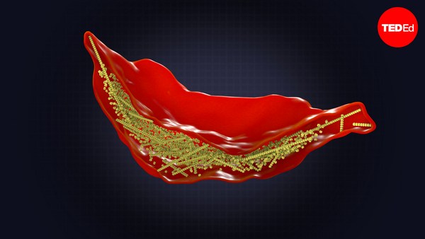
And it took us a long time to understand where they came from. They come from these cells. These blue and red cells are called doublecortin-positive cells. All of you have them in your brain. They represent four percent of your cortical brain cells. They have a very important role during the development stage. When you were fetuses, they helped your brain to fold itself. But why do they stay in your head? This, we don't know. We think that they may participate in brain repair because we find them in higher concentration close to brain lesions. But it's not so sure. But there is one clear thing -- that from these cells, we got our stem cell culture. And we were in front of a potential new source of cells to repair the brain. And we had to prove this.
Het duurde een tijd voor we begrepen waar ze vandaan kwamen. Ze komen uit deze cellen: die blauwe en rode cellen zijn doublecortin-positieve cellen. Je hebt ze allemaal in je brein. Ze beslaan 4 procent van je corticale hersencellen. Ze spelen een belangrijke rol in de ontwikkeling. Toen jullie nog foetussen waren, hielpen ze het brein om zich in te vouwen. Maar, waarom blijven ze in je hoofd? Dat weten we niet. We denken dat ze het brein helpen te herstellen omdat we ze in hogere concentraties vinden bij hersenlaesies. Dit is echter niet zeker. Eén ding is zeker: uit deze cellen ontstaat onze stamcelcultuur. We stonden voor een potentieel nieuwe bron van cellen die het brein kunnen herstellen. We moesten het bewijzen,
So to prove it, we decided to design an experimental paradigm. The idea was to biopsy a piece of brain in a non-eloquent area of the brain, and then to culture the cells exactly the way Jean-François did it in his lab. And then label them, to put color in them in order to be able to track them in the brain. And the last step was to re-implant them in the same individual. We call these autologous grafts -- autografts.
en daarom hebben we een experimenteel paradigma opgezet. We wilden een biopsie nemen van hersenweefsel dat niet in een spraakgebied ligt, en die cellen op kweek zetten, precies zoals Jean-François in zijn lab deed. We labelden ze daarna met kleur zodat we ze konden volgen in het brein. Tot slot wilden we ze herimplanteren in hetzelfde individu. We noemen dit autogene of autotransplantatie.
So the first question we had, "What will happen if we re-implant these cells in a normal brain, and what will happen if we re-implant the same cells in a lesioned brain?" Thanks to the help of professor Eric Rouiller, we worked with monkeys.
Onze eerste vraag was: wat gebeurt er als we deze cellen in een normaal brein herimplanteren, en wat als we de dat doen in een beschadigd brein? Dankzij de hulp van professor Eric Rouiller konden we met apen werken.
So in the first-case scenario, we re-implanted the cells in the normal brain and what we saw is that they completely disappeared after a few weeks, as if they were taken from the brain, they go back home, the space is already busy, they are not needed there, so they disappear.
Dus in het eerste scenario herimplanteerden we de cellen in een normaal brein. We zagen dat ze na enkele weken geheel verdwenen waren, alsof ze waren verwijderd. Ze gaan naar hun gebied en als er geen plaats is en ze niet nodig zijn, verdwijnen ze.
In the second-case scenario, we performed the lesion, we re-implanted exactly the same cells, and in this case, the cells remained -- and they became mature neurons. And that's the image of what we could observe under the microscope. Those are the cells that were re-implanted. And the proof they carry, these little spots, those are the cells that we've labeled in vitro, when they were in culture.
In het tweede scenario hebben we een laesie gemaakt en daarna dezelfde cellen geherimplanteerd en nu bleven de cellen ter plaatse en ze werden volgroeide neuronen. Dit zagen we onder de microscoop, dit zijn de cellen die we herimplanteerden, en het bewijs: deze kleine vlekjes zijn de cellen die we in vitro labelden, toen ze op kweek stonden.
But we could not stop here, of course. Do these cells also help a monkey to recover after a lesion? So for that, we trained monkeys to perform a manual dexterity task. They had to retrieve food pellets from a tray. They were very good at it. And when they had reached a plateau of performance, we did a lesion in the motor cortex corresponding to the hand motion. So the monkeys were plegic, they could not move their hand anymore. And exactly the same as humans would do, they spontaneously recovered to a certain extent, exactly the same as after a stroke. Patients are completely plegic, and then they try to recover due to a brain plasticity mechanism, they recover to a certain extent, exactly the same for the monkey.
Maar, we waren nog niet klaar. Kunnen deze cellen een aap helpen herstellen na een laesie? We trainden de apen in een manuele taak. Ze moesten korreltjes voedsel van een blad afhalen. Ze waren er erg goed in. Toen ze een bepaald niveau hadden bereikt, maakten we een laesie in de motorische cortex waar de beweging van de hand zit. Dat verlamde de apen, ze konden hun hand niet meer bewegen. Net als mensen, herstelden ze vanzelf tot een bepaald niveau, net als na een beroerte. Patienten die geheel verlamd zijn proberen gedeeltelijk te herstellen, dankzij de plasticiteit van het brein, en zo ging het ook bij de aap.
So when we were sure that the monkey had reached his plateau of spontaneous recovery, we implanted his own cells. So on the left side, you see the monkey that has spontaneously recovered. He's at about 40 to 50 percent of his previous performance before the lesion. He's not so accurate, not so quick. And look now when we re-implant the cells: Two months after re-implantation, the same individual.
Toen we zeker wisten dat de aap het niveau had bereikt van spontaan herstel, implanteerden we zijn eigen cellen. Links ziet u de aap die spontaan hersteld is. Hij zit op 40 à 50 procent van zijn vermogen van voor de laesie. Hij is niet zo accuraat en snel. En kijk, toen we cellen herimplanteerden: dezelfde aap twee maanden na herimplantatie!
(Applause)
(Applaus)
It was also very exciting results for us, I tell you. Since that time, we've understood much more about these cells. We know that we can cryopreserve them, we can use them later on. We know that we can apply them in other neuropathological models, like Parkinson's disease, for example. But our dream is still to implant them in humans. And I really hope that I'll be able to show you soon that the human brain is giving us the tools to repair itself.
Voor ons waren het ook opwindende resultaten. Sindsdien begrijpen we veel meer van deze cellen. We weten dat we ze kunnen cryopreserveren voor later gebruik. We kunnen ze gebruiken bij andere neurologische aandoeningen, zoals de ziekte van Parkinson. Het is nog steeds onze droom ze in mensen te implanteren. Ik hoop dat ik jullie snel kan laten zien dat het menselijke brein ons het gereedschap geeft zichzelf te herstellen.
Thank you.
Dank je wel.
(Applause)
(Applaus)
Bruno Giussani: Jocelyne, this is amazing, and I'm sure that right now, there are several dozen people in the audience, possibly even a majority, who are thinking, "I know somebody who can use this." I do, in any case. And of course the question is, what are the biggest obstacles before you can go into human clinical trials?
Bruno Giussani: Jocelyne, dit is geweldig. Er zijn vast mensen in het publiek misschien de meerderheid, die denken: "Ik ken iemand die dit kan gebruiken." Ik in elk geval wel. De vraag is nu: wat staat er in de weg voordat we dit op mensen kunnen testen?
Jocelyne Bloch: The biggest obstacles are regulations. (Laughs) So, from these exciting results, you need to fill out about two kilograms of papers and forms to be able to go through these kind of trials.
Jocelyne Bloch: vooral regeltjes. (Lacht) Voor deze prachtige resultaten moet je twee kilo papier en formulieren invullen om met dit soort testen te starten.
BG: Which is understandable, the brain is delicate, etc.
BG: Begrijpelijk, want het brein is kwetsbaar, enz.
JB: Yes, it is, but it takes a long time and a lot of patience and almost a professional team to do it, you know?
JB: Ja, dat klopt, maar het kost veel tijd en geduld en bijna een heel team om het uit te voeren.
BG: If you project yourself -- having done the research and having tried to get permission to start the trials, if you project yourself out in time, how many years before somebody gets into a hospital and this therapy is available?
BG: Kijk eens vooruit: je hebt onderzoek gedaan, en geprobeerd toestemming te krijgen om testen op te starten. Als je dan in de toekomst kijkt: hoeveel jaar dan tot iemand in het ziekenhuis komt en deze therapie beschikbaar is?
JB: So, it's very difficult to say. It depends, first, on the approval of the trial. Will the regulation allow us to do it soon? And then, you have to perform this kind of study in a small group of patients. So it takes, already, a long time to select the patients, do the treatment and evaluate if it's useful to do this kind of treatment. And then you have to deploy this to a multicentric trial. You have to really prove first that it's useful before offering this treatment up for everybody.
JB: Dat is moeilijk te zeggen. Eerst moet het onderzoek worden toegestaan. Zal de wet ons toestaan dat gauw te doen? Dan moet je dit onderzoek uitvoeren met een kleine patiëntengroep. Het duurt lang om patiënten te selecteren, de behandeling te doen en het nut ervan te evalueren. Het onderzoek moet dan uitgebreid worden naar andere hospitalen en andere dokters. Eerst moet je het nut bewijzen, voor je het aan iedereen kunt aanbieden.
BG: And safe, of course. JB: Of course.
BG: En veilig, natuurlijk. JB: Uiteraard.
BG: Jocelyne, thank you for coming to TED and sharing this. BG: Thank you.
BG: Jocelyne, bedankt voor je komst en dit met ons te delen. BG: Dank je.
(Applause)
(Applaus)