You know, cadaver dissection is the traditional way of learning human anatomy. For students, it's quite an experience, but for a school, it could be very difficult or expensive to maintain. So we learned the majority of anatomic classes taught, they do not have a cadaver dissection lab. Maybe those reasons, or depending on where you are, cadavers may not be easily available.
אתם יודעים, נתיחת גופות היא הדרך המסורתית ללימוד האנטומיה האנושית. מבחינת הסטודנטים זו חוויה רצינית, אך מבחינת בית הספר, זה עלול להיות קשה מאד או יקר מבחינת תחזוקה. נוכחנו לדעת שרוב שיעורי האנטומיה מועברים, בלי מעבדה לביתור גופות. אולי מהסיבות האלה, או לפי המיקום שלכם, לא תמיד קל להשיג גופות.
So to address this, we developed with a Dr. Brown in Stanford: virtual dissection table. So we call this Anatomage Table. So with this Anatomage Table, students can experience the dissection without a human cadaver. And the table form is important, and since it's touch-interactive, just like the way they do dissections in the lab, or furthermore just the way a surgeon operates on a patient you can literally interact with your table. Our digital body is one-to-one life size, so this is exactly the way students will see the real anatomy.
כדי לטפל בזה, פיתחנו יחד עם דוקטור בראון בסטנפורד שולחן ניתוח וירטואלי. אנחנו קוראים לזה שולחן אנטומג'. אז עם השולחן הזה, סטודנטים יכולים לחוות את הניתוח בלי גופה אנושית. וצורת השולחן היא חשובה, ומאחר והוא רגיש למגע, בדיוק כמו הדרך בה מנתחים במעבדה, או בדיוק בדרך בה מנתח מטפל בחולה אתם ממש יכולים לתקשר עם השולחן. הגוף הדיגיטלי הוא בגודל אמיתי, זו בדיוק הדרך בה הסטודנטים יראו את האנטומיה האמיתית.
I'm going to do some demonstrations. As you can see, I use my finger to interact with my digital body. I'm going to do some cuts. I can cut any way I want to, so I cut right here. Then it's going to show inside. And I can change my cut to see different parts. Maybe I can cut there, see the brain, and I can change my cut. You can see some internal organs. So we call this the slicer mode. OK, I'm going to do another cut. Right there. This shows a lot of internal structures. So if I want to see the back side, I can flip and see from behind. Like this. So if these images are uncomfortable to you or disturbing to you, that means we did the right job. So our doctors said these are eye candies.
אני עומד להציג כמה הדגמות. כמו שאתם רואים, אני משתמש באצבעי כדי להפעיל את שולחן הגוף הדיגיטלי. אני עומד לבצע כמה חיתוכים. אני יכול לחתוך בכל דרך שארצה, אז אני חותך בדיוק פה. כעת זה יציג מראה פנימי. ואני יכול לשנות את החתך שלי כדי לראות חלקים שונים. אולי אני יכול לחתוך שם, לראות את המוח, ואני יכול לשנות את החיתוך שלי. אתם יכולים לראות כמה אברים פנימיים. אז קוראים לזה מצב חיתוך. אוקיי, אני אבצע עוד חיתוך. בדיוק שם. זה מראה הרבה מבנים פנימיים. אז אם אני רוצה לראות את הצד האחורי, אני יכול להפוך ולראות מאחור. ככה. אז אם התמונות האלה הן גורמות לכם אי נוחות או מטרידות אתכם, זה אומר שעשינו עבודה טובה. הרופאים שלנו אמרו שזו חגיגה לעיניים.
So instead of just butchering the body, I'd like to do more clinically meaningful dissections. What I'm going to do is I'm going to peel off all the skin, muscles and bones, just to see a few internal organs. Right here. Let's say I'm going to cut the liver right here. OK. Let's say I'm interested in looking at the heart. I'm going to do some surgery here. I'm going to cut some veins, arteries. Oops! ...
אז במקום סתם לבתר את הגוף, הייתי רוצה לעשות חיתוכים בעלי משמעות קלינית גדולה יותר. מה שאני עומד לעשות הוא לקלף את העור, השרירים והעצמות, כדי לראות כמה איברים פנימיים. בדיוק כאן. נניח שאני רוצה לחתוך את הכבד בדיוק כאן. בסדר. נניח שאני רוצה להביט בלב. אני אנתח פה מעט. אני אחתוך כמה ורידים, עורקים. אופס!...
You don't want to hear "oops" in real surgery. (Laughter) But fortunately, our digital man has "undo." (Laughter) Okay.
אתם לא רוצים לשמוע "אופס" בניתוח אמיתי. (צחוק) אבל למזלנו, לאיש הדיגיטלי שלנו יש "בטל פעולה." (צחוק) אוקיי.
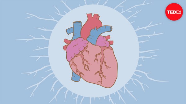
All right then. Let me zoom in. I'm going to make a cut right there. And then you can see the inside of the heart. You can see the atrium and the ventricles, how blood flows to our arteries and veins. Just like this, students can isolate anybody and dissect any way you want to. It doesn't have to be always dissection. Since it's digital, we can do reverse dissection. So let me show you, I'm going to start with the skeletal structure, and I can add a few internal organs. Yep. Maybe I can add quickly this way. And I can build muscles gradually, just like that. We can see tendons and muscles. Wish I could build my muscle this fast. (Laughter) And this is another way to learn anatomy.
אז בסדר. אבצע כאן תקריב. אני עומד לבצע חתך בדיוק שם. ואתם יכולים לראות את תוך הלב. אתם יכולים לראות את העליות והחדרים, איך הדם זורם לעורקים ולוורידים שלנו. בדיוק ככה, הסטודנט יכול לבודד כל דבר ולחתוך בכל דרך שירצה. זה לא חייב להיות חיתוכים כל הזמן. מאחר וזה דיגיטלי, אנחנו יכולים לעשות ניתוח הפוך. אז תנו לי להראות לכם, אני אתחיל עם מבנה השלד, אני יכול להוסיף כמה אברים פנימיים. כן. אולי אני יכול להוסיף בזריזות ככה. ואני יכול לבנות שרירים בהדרגה. בדיוק ככה. אנחנו יכולים לראות רקמות חיבור ושרירים. הלוואי ויכולתי לבנות את השרירים שלי כל כך מהר. (צחוק) וזו דרך נוספת ללמוד אנטומיה.
Another thing I can show you is, more often than not, doctors get to meet patients in X-ray form. So, Anatomage Table shows exactly how the anatomy will appear in X-ray. You can also interact with your X-ray, and also if you want, you can compare with how anatomy would appear in X-ray, too. So when you are done, just bring back the body and then it's ready for another session. It looks like our table also can transform gender, too. It's a female now.
דבר נוסף שאני יכול להראות לכם הוא, שהרבה פעמים, הרופא פוגש את החולה שלו בצורת צילום רנטגן. אז, השולחן מראה בדיוק איך האנטומיה תיראה בצילום רנטגן. אתם יכולים גם לעבוד עם צילומי רנטגן, ואם אתם רוצים, אתם גם יכולים להשוות מול איך שהאנטומיה היתה נראית גם בצילום רנטגן. אז כשאתם מסיימים, פשוט תחזירו את הגוף ואז הוא מוכן לאדם נוסף. זה נראה כאילו השולחן שלנו יכול גם לשנות מין. זו אישה עכשיו.
So this is Anatomage Table. Thank you. (Applause)
אז זה שולחן האנטומאג'. תודה לכם. (מחיאות כפיים)