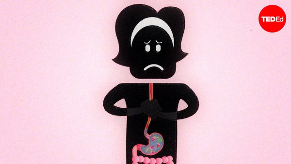
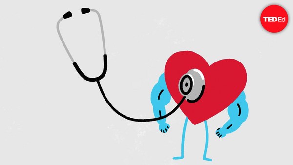
For most of history, humans had no idea what purpose the heart served. In fact, the organ so confused Leonardo da Vinci, that he gave up studying it. Although everyone could feel their own heart beating, it wasn't always clear what each thump was achieving. Now we know that the heart pumps blood. But that fact wasn't always obvious, because if a heart was exposed or taken out, the body would perish quickly. It's also impossible to see through the blood vessels, and even if that were possible, the blood itself is opaque, making it difficult to see the heart valves working. Even in the 21st century, only a few people in surgery teams have actually seen a working heart. Internet searches for heart function, point to crude models, diagrams or animations that don't really show how it works. It's as if there has been a centuries old conspiracy amongst teachers and students to accept that heart function cannot be demonstrated. Meaning that the next best thing is simply to cut it open and label the parts. That way students might not fully grasp the way it works, but can superficially understand it, learning such concepts as the heart is a four-chambered organ, or potentially misleading statements like, mammals have a dual-circulation: one with blood going to the lungs and back, and another to the body and back. In reality, mammals have a figure-eight circulation. Blood goes from one heart pump to the lungs, back to the second heart pump, which sends it to the body, and then back to the first pump. That's an important difference because it marks two completely different morphologies. This confusion makes many students wary of the heart in biology lessons, thinking it signals an intimidating subject full of complicated names and diagrams. Only those who end up studying medicine compeltely understand how it all actually works. That's when its functions become apparent as medics get to observe the motion of the heart's valves. So, let's imagine you're a medic for a day. What you'll need to get started is a whole fresh heart, like one from a sheep or pig. Immerse this heart in water and you'll see that it doesn't pump when squeezed by hand. That's because water doesn't enter the heart cleanly enough for the pumping mechanism to work. We can solve this problem in an extraordinarly simple way. Simply identify the two atria and cut them off, trimming them down to the tops of the ventricles. This makes the heart look less complicated because the atria have several incoming veins attached. So without them there, the only vessels remaining are the two major heart arteries: the aorta and pulmonary artery, which rise like white columns from between the ventricles. It looks -- and really is -- very simple. If you run water into the right ventricle from a tap (the left also works, but less spectacularly), you'll see that the ventricular valve tries to close against the incoming stream. And then ventricle inflates with water. Squeeze the ventricle and a stream of water squirts out of the pulmonary artery. The ventricular valves, called the tricuspid in the right ventricle and the mitral in the left, can be seen through the clear water opening and closing like parachutes as the ventricle is rhythmically squeezed. This flow of water mimics the flow of blood in life. The valves are completely efficient. You'll notice they don't leak at all when the ventricles are squeezed. Over time, they also close against each other with very little wear and tear, which explains how this mechanism continues to work seamlessly for more than 2 billion beats a heart gives in its lifetime. Now, anyone studying the heart can hold one in their hands, make it pump for real and watch the action unfold. So place your hand above your own and feel its rhymic beat. Understanding how this dependable inner pump works gives new resonance to the feeling you get when you run a race, drink too much caffeine or catch the eye of the one you love.
Bardzo długo ludzie nie mieli pojęcia, do czego służy serce. Ten organ do tego stopnia zdezorientował Leonarda da Vinci, że porzucił badania nad nim. Choć każdy może poczuć bicie swojego serca, jego cel nie zawsze był jasne. Teraz wiemy, że serce pompuje krew. Ale to nie zawsze było takie oczywiste, bo kiedy serce było odsłonięte czy wyjęte, ciało szybko umierało. Nie można też patrzeć przez żyły, ale nawet gdyby było to możliwe, krew jest nieprzejrzysta, przez co trudno dostrzec pracę zastawek. Nawet w XXI wieku bardzo niewielu chirurgów widziało pracujące serce na żywo. Wyszukiwarki internetowe w sprawie serca odsyłają do prymitywnych modeli, diagramów albo animacji, które nie pokazują, jak naprawdę działa serce. To tak, jakby odwieczna konspiracja między nauczycielami i studentami wymuszała akceptację, że funkcjonowania serca nie da się zademonstrować. Można tylko rozciąć serce i oznakować jego części. Studenci nie załapią do końca jak pracuje, ale powierzchownie mogą się dowiedzieć, że serce jest organem czterokomorowym, albo przyjmą potencjalnie mylące tezy, jak na przykład, że ssaki mają dwa obiegi krwionośne, jeden biegnący do płuc i z powrotem, drugi do ciała i z powrotem. W rzeczywistości układ krwionośny ssaków jest jak leniwa ósemka. Krew przepływa z prawej części serca do płuc, wraca do lewej części serca, która wysyła ją dalej, do ciała, a stamtąd krew wraca do prawej części serca. To ważna różnica, bo to oznacza dwie zupełnie różne morfologie. To zamieszanie sprawia, że wielu uczniów dość oględnie traktuje serce podczas lekcji biologii, myśląc, że to zwiastuje coś strasznie trudnego, na pewno z mnóstwem skomplikowanych nazw i diagramów. Tylko studenci medycyny rozumieją do końca, jak to wszystko działa. Funkcje serca stają się oczywiste wtedy, gdy medycy muszą obserwować ruchy zastawek. Wyobraźmy sobie, że na dziś jesteś studentem medycyny. Na początek będziesz potrzebować całego, świeżego serca, na przykład od owcy albo świni. Zanurz to serce w wodzie, a zauważysz, że ściskane dłonią wcale nie pompuje. Dzieje się tak, bo woda nie wpływa do serca na tyle czysto, żeby mechanizm pompujący mógł zacząć pracować. Możemy rozwiązać ten problem w nadzwyczaj prosty sposób. Znajdź dwa przedsionki i odetnij je, zmniejszając je aż do szczytu komory. To sprawia, że serce wygląda prościej, bo do przedsionków są przytwierdzone żyły. Bez nich jedyne pozostałe naczynia krwionośne, to dwie najważniejsze tętnice: aorta i tętnica płucna, które wystają jak kolumny spomiędzy komór. To wygląda, i jest, naprawdę proste. Jeżeli do prawej komory nalejesz wody z kranu, (lewa też zadziała, ale mniej spektakularnie) zobaczysz, że zastawka komory próbuje się zamknąć, żeby nie wpuścić strumienia wody. A kiedy komora napełni się wodą ściśnij ją, a strumień wody wytryśnie z tętnicy płucnej. Zastawki komór zwane w prawej komorze trójdzielną, a w lewej mitralną, są widoczne w czystej wodzie, otwierające się i zamykające jak spadochron, kiedy komora jest rytmicznie ściskana. Ten przepływ wody imituje prawdziwy przepływ krwi. Zastawki są bardzo efektywne. Zauważ, że w ogóle nie przeciekają, kiedy ściska się komorę. Z czasem zaczynają też zamykać się na zmianę, nie zużywając się ani nie niszcząc, co tłumaczy, jak serce działa bez przerwy przez ponad 2 miliardy uderzeń w ciągu twojego życia. Teraz każdy adept serca może potrzymać je w ręku, sprawić, że zacznie pompować i śledzić rozwój akcji. Połóż dłoń na sercu i spróbuj wyczuć jego rytmiczne uderzenia. Zrozumienie działania tej niezawodnej pompy, nadaje nowy wydźwięk uczuciu, którego doświadczasz podczas wyścigu, gdy wypijesz za dużo kofeiny albo zobaczysz swoją miłość.