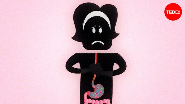
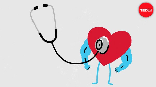
For most of history, humans had no idea what purpose the heart served. In fact, the organ so confused Leonardo da Vinci, that he gave up studying it. Although everyone could feel their own heart beating, it wasn't always clear what each thump was achieving. Now we know that the heart pumps blood. But that fact wasn't always obvious, because if a heart was exposed or taken out, the body would perish quickly. It's also impossible to see through the blood vessels, and even if that were possible, the blood itself is opaque, making it difficult to see the heart valves working. Even in the 21st century, only a few people in surgery teams have actually seen a working heart. Internet searches for heart function, point to crude models, diagrams or animations that don't really show how it works. It's as if there has been a centuries old conspiracy amongst teachers and students to accept that heart function cannot be demonstrated. Meaning that the next best thing is simply to cut it open and label the parts. That way students might not fully grasp the way it works, but can superficially understand it, learning such concepts as the heart is a four-chambered organ, or potentially misleading statements like, mammals have a dual-circulation: one with blood going to the lungs and back, and another to the body and back. In reality, mammals have a figure-eight circulation. Blood goes from one heart pump to the lungs, back to the second heart pump, which sends it to the body, and then back to the first pump. That's an important difference because it marks two completely different morphologies. This confusion makes many students wary of the heart in biology lessons, thinking it signals an intimidating subject full of complicated names and diagrams. Only those who end up studying medicine compeltely understand how it all actually works. That's when its functions become apparent as medics get to observe the motion of the heart's valves. So, let's imagine you're a medic for a day. What you'll need to get started is a whole fresh heart, like one from a sheep or pig. Immerse this heart in water and you'll see that it doesn't pump when squeezed by hand. That's because water doesn't enter the heart cleanly enough for the pumping mechanism to work. We can solve this problem in an extraordinarly simple way. Simply identify the two atria and cut them off, trimming them down to the tops of the ventricles. This makes the heart look less complicated because the atria have several incoming veins attached. So without them there, the only vessels remaining are the two major heart arteries: the aorta and pulmonary artery, which rise like white columns from between the ventricles. It looks -- and really is -- very simple. If you run water into the right ventricle from a tap (the left also works, but less spectacularly), you'll see that the ventricular valve tries to close against the incoming stream. And then ventricle inflates with water. Squeeze the ventricle and a stream of water squirts out of the pulmonary artery. The ventricular valves, called the tricuspid in the right ventricle and the mitral in the left, can be seen through the clear water opening and closing like parachutes as the ventricle is rhythmically squeezed. This flow of water mimics the flow of blood in life. The valves are completely efficient. You'll notice they don't leak at all when the ventricles are squeezed. Over time, they also close against each other with very little wear and tear, which explains how this mechanism continues to work seamlessly for more than 2 billion beats a heart gives in its lifetime. Now, anyone studying the heart can hold one in their hands, make it pump for real and watch the action unfold. So place your hand above your own and feel its rhymic beat. Understanding how this dependable inner pump works gives new resonance to the feeling you get when you run a race, drink too much caffeine or catch the eye of the one you love.
역사의 대부분에서, 인간은 심장이 제공하는 목적을 전혀 몰랐어요. 사실, 그 기관은 리오나르도 다 빈치를 너무 혼란케해서 그는 그 공부하기를 포기했어요. 모든 사람이 그 자신의 심작박동을 느낄 수 있지만, 각 박동이 성취하는게 뭔지는 늘 분명하지 않았지요. 이제 우리는 심장이 피를 뿜는다는걸 압니다. 하지만, 그 사실은 항상 명백하지 않았죠. 왜냐면 만일 심장이 드러났거나 떼어내지면, 그 몸은 빨리 죽을것이기 때문이죠. 그건 또한 혈관을 통해서 보기가 불가능하고, 심지어 그게 가능하다 해도, 그 피 자체는 불통명해서, 심장의 판막이 작동하는지 보기는 어렵게 하죠. 21세기에도. 수술팀의 단 몇명만이 움직이고 있는 심장을 보았어요. 인터넷은 심장이 박동하는 방법을 실제로 보여주지 않는 심장기능, 한 점에서 천연모델의, 다이어그램이나 에니매이션을 조사합니다. 그건 마치 선생님들과 학생들 사이에서 심장기능은 전시될 수 없다는 걸 인정하는 음모가 100년이나 지속된 것 같습니다. 그게 의미하는 것은 다음에 가장 최고인것은, 간단히 심장을 절개개방해 각 부분에 이름을 짓는다는 거죠. 그러면 학생들은 심장이 작동하는 법을 완전히 이해할 수 없지만 그걸 피상적으로 이해할 수 있으니까요. 그 같은 개념을 배우기를 심장이 -4심실 기관이라고 할 수 있거나, 잠재적으로 혼동시키는 진술을 할 수 있어요. 포유동물은 2중- 순환기를 지니고 있다고요: 피를 가지고 폐로 갔다가 돌아오는 순환기 하나와, 몸으로 갔다가 돌아오는 다는 또 다른 순환기라구요. 실제로는, 포유동물 8자 모양의 순환기를 가지고 있어요. 피는 심장박동 하나에서 폐로 갔다가, 두번째 심장박동으로 돌아갑니다. 즉 몸으로 보내지고, 다시 첫번째 박동으로 돌아갑니다. 그건 중요하게 다른점입니다. 그건 두개의 완전히 다른 형태을 표시하기 때문이죠. 이 혼란은 많은 학생들을 생물학 수업에서 심장에 대해 피곤하게 만들죠, 그건 복잡한 이름들과 도형으로 가득찬 두렵게만드는 과목을 표시한다고 생각하며 말이죠. 나중에 의약을 공부하게 되는 학생들만이 그게 전부 작용하는 방법을 완전히 이해합니다. 그건 기능이 명백해질 때 입니다. 심장 판막의 움직임을 관찰하는 수련의사로서 말이죠. 그러니, 여러분이 하루동안 수련의사라고 상상해 보죠. 시작하기위해 필요한 것은 생생한 심장전체입니다. 양이나 돼지에서 나온것 같은거죠. 이 심장을 물속에 담그고 손으로 압착시키면 심장이 뛰지 않는걸 보실겁니다. 물이 충분히 깨끗하게 들어가지 않기 때문이죠. 심장에 박동의 메커니즘이 작용하기 위해서요. 우리는 극도로 간단한 식으로 이 문제를 풀 수 있어요. 단순히 심방을 식별해서 그걸 절단하고, 그걸 심실의 위로 잘라갑니다. 이건 심장이 덜 복잡하게 보이게 합니다. 왜냐면 그 심방은 몇개의 도입혈관이 붙었거든요. 그것들이 거기 없으면, 오직 남아있는 혈관은 두개의 주요 심장동맥이에요: 대동맥과 폐 대동맥, 즉, 심실사이에서 하얀 기둥처럼 올라오는 거죠. 그렇게 보이고 -또 실제로 그래요-아주 간단하죠. 만일 꼭지에서 오른쪽 심실로 물을 흘러보내면 (왼쪽도 작용하지만, 덜 극적이에요) 심실 판막이 들어오는 물줄기에 역행해서 닫으려고 하는 걸 보실거에요. 그러면 심실은 물로 팽창해요. 심실을 압착하면 그 물줄기는 폐 동맥의 밖으로 분출해요. 오른 쪽 심실에서 삼첨판이라 불리는 심실의 판막과, 왼쪽의 승모판은 낙하산처럼 열고 닫는것을 투명한 물을 통해 볼 수 있는데 심실이 리드미컬하게 압착되었기 때문이죠. 이 물의 흐름은 생명에서 피의 흐름을 흉내냅니다. 그 판막은 완전히 효율적이죠. 여러분은 심실이 압착되었을 때 새지 않음을 아실 겁니다. 시간이 지나면, 그것들은 또 서로에 대항해 닫습니다 아주 사소한 손실을 보이면서요. 즉 이 체계는 계속 실수없이 하나의 심장이 그 생에서 20억번의 박동을 하는지의 방법을 설명하는 거죠. 심장을 공부하는 그 어떤 학생이라도 심장을 손에 들고, 그 박동을 실제로 만들고 그 행위가 전개되는 것을 볼 수 있어요. 그러니 여러분의 손을 심장위에 엊고 그 리듬감있는 비트를 느끼세요. 이 믿을만한 내면의 펌프가 작용하는 방법을 아는것은 여러분이 느끼는것에 새로운 울림을 드립니다. 여러분이 달리기 경주를 하거나, 카페인을 너무 많이 마시거나, 여러분이 사랑하는것을 눈에 담았을때 말이죠.