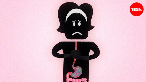
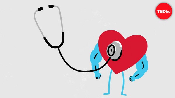
For most of history, humans had no idea what purpose the heart served. In fact, the organ so confused Leonardo da Vinci, that he gave up studying it. Although everyone could feel their own heart beating, it wasn't always clear what each thump was achieving. Now we know that the heart pumps blood. But that fact wasn't always obvious, because if a heart was exposed or taken out, the body would perish quickly. It's also impossible to see through the blood vessels, and even if that were possible, the blood itself is opaque, making it difficult to see the heart valves working. Even in the 21st century, only a few people in surgery teams have actually seen a working heart. Internet searches for heart function, point to crude models, diagrams or animations that don't really show how it works. It's as if there has been a centuries old conspiracy amongst teachers and students to accept that heart function cannot be demonstrated. Meaning that the next best thing is simply to cut it open and label the parts. That way students might not fully grasp the way it works, but can superficially understand it, learning such concepts as the heart is a four-chambered organ, or potentially misleading statements like, mammals have a dual-circulation: one with blood going to the lungs and back, and another to the body and back. In reality, mammals have a figure-eight circulation. Blood goes from one heart pump to the lungs, back to the second heart pump, which sends it to the body, and then back to the first pump. That's an important difference because it marks two completely different morphologies. This confusion makes many students wary of the heart in biology lessons, thinking it signals an intimidating subject full of complicated names and diagrams. Only those who end up studying medicine compeltely understand how it all actually works. That's when its functions become apparent as medics get to observe the motion of the heart's valves. So, let's imagine you're a medic for a day. What you'll need to get started is a whole fresh heart, like one from a sheep or pig. Immerse this heart in water and you'll see that it doesn't pump when squeezed by hand. That's because water doesn't enter the heart cleanly enough for the pumping mechanism to work. We can solve this problem in an extraordinarly simple way. Simply identify the two atria and cut them off, trimming them down to the tops of the ventricles. This makes the heart look less complicated because the atria have several incoming veins attached. So without them there, the only vessels remaining are the two major heart arteries: the aorta and pulmonary artery, which rise like white columns from between the ventricles. It looks -- and really is -- very simple. If you run water into the right ventricle from a tap (the left also works, but less spectacularly), you'll see that the ventricular valve tries to close against the incoming stream. And then ventricle inflates with water. Squeeze the ventricle and a stream of water squirts out of the pulmonary artery. The ventricular valves, called the tricuspid in the right ventricle and the mitral in the left, can be seen through the clear water opening and closing like parachutes as the ventricle is rhythmically squeezed. This flow of water mimics the flow of blood in life. The valves are completely efficient. You'll notice they don't leak at all when the ventricles are squeezed. Over time, they also close against each other with very little wear and tear, which explains how this mechanism continues to work seamlessly for more than 2 billion beats a heart gives in its lifetime. Now, anyone studying the heart can hold one in their hands, make it pump for real and watch the action unfold. So place your hand above your own and feel its rhymic beat. Understanding how this dependable inner pump works gives new resonance to the feeling you get when you run a race, drink too much caffeine or catch the eye of the one you love.
歴史の大部分のうち 人類は心臓の役割を全く理解する事ができませんでした あのダヴィンチでさえも 心臓の分かりにくさに 勉強する事を諦めたほどでした 誰もが自分の心臓の鼓動を感じる事は出来ましたが その鼓動ひとつひとつが何を行っているのか ハッキリとしませんでした 今では心臓は血液を送り出すもの と分かりますが その事実は常に明らかではありませんでした それは 心臓をむき出しにしたり 取り出したりすると 身体がすぐに滅びてしまうからです また血管を通して内部を見る事もできません たとえ出来たとしても 血液が不透明であるために 心臓の弁の動きを見る事はとても困難です 21世紀であっても 外科医チームにいる一部の人しか 実際に動いている心臓を見たことはありません インターネットで心臓の働きを検索すると 大雑把な作りの模型や図形 また正しく描かれていない アニメーションなどにたどり着きます まるで心臓の動きを学ぶ事は不可能である ということを 受け入れてしまおう という考えが 教師と生徒の間で 何百年も 続いて来たかのようです 次に出来る事は 単に心臓を開けてみて それぞれの部分に 名前をつけることです 心臓の働きを完全に掴む事は 出来ないかもしれませんが 表面的に理解をする事は出来ます そして 心臓は四つに仕切られている と言う事などを学ぶができます 同時に 間違った解釈に繋がる事もあります 例えば ほ乳類の体には 二重の血液循環がある —肺を巡るものと、体を巡るものー と言うものなどです 実際の所 ほ乳類の体には 八の字型の血液循環があります 血液は心臓のひとつのポンプから肺に送り出され 二つ目のポンプに戻り そこから体全体に送り出され そしてまた 最初のポンプに戻ってゆきます それが重要な違いです この二つは全く異なった構造です この様に分かりにくい事から 多くの学生が 生物で心臓について学ぶ時に慎重になります 複雑な名前や図形でいっぱいの難しい内容であると 考えてしまうのです 最終的に 医学を勉強する人達だけが 心臓の働きを完全に理解するのです 医学生達は 心臓の弁の動きを観察する事で その働きをより明確に理解する事ができます さて 今日はあなたが 医学生であると想像しましょう まず最初に必要なのは 取り出したばかりの心臓 羊か豚のものが良いでしょう 心臓を水に沈めてみましょう すると 心臓を手で握っても 水を送り出さない事が分かるでしょう これは 心臓のポンプが働くほど 水がうまく心臓に入って行かないからです この事は 驚く程簡単な方法で 解決する事が出来ます それには 二つの心房を見つけ出し 切除し 心室の一番上まで 切り取ってしまう事です これで心臓はもう少し見やすくなるでしょう 心房にはいくつかの静脈がついているため それらの血管がなくなってしまえば 残りの血管は 二つの重要な動脈だけになります つまり 大動脈と肺動脈です これらの動脈は二つの心室の間から 円柱のように伸びています 大変シンプルな見た目と作りです 右心室に水道水を流し入れてみましょう (左心室でも大丈夫ですが 右心室ほど効果がありません) すると心室の弁が 入ってくる水の流れに対抗して 閉まろうとするのが分かるでしょう 水が入った心室はやがて膨張します 手で握りしめると 水が 肺動脈から 飛び出すでしょう 右心室の弁は 三尖弁 左心室の弁は 僧帽弁 とそれぞれ 呼ばれています 透明な水を通して見ると 一定のリズムで心室が握られたとき それらの弁がパラシュートのように 開閉するのが分かるでしょう この流れは実際の血液の流れと似ています これらの弁の効果は抜群です 心室が握られている時も 水が一切漏れない事に気づくでしょう 時間とともにほぼ擦り切れる事もなく 閉まり続けるのです そのおかげで 一生のうち 約20億回以上という数の鼓動を 心臓は 絶え間なく 打ち続ける事ができるのです 心臓を勉強している人なら 実際に心臓を手に取り 動かしてみることで その働きが明らかになるのを見る事ができます あなたも自分の心臓に手を当てて 心臓のリズミカルな鼓動を感じてみましょう この頼もしいポンプが体内で どう働くのかを理解すると レースで競争したり カフェインを沢山飲んだり あの子と目が合ったり そんな時に感じるドキドキが より意味の深いものに感じるでしょう