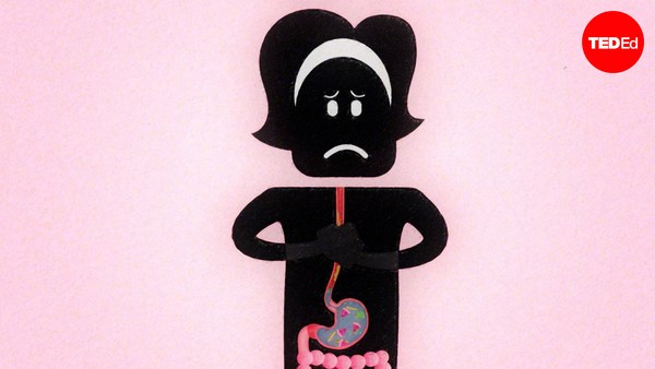
لفترة طويلة من التاريخ، لم يتوفر لدى البشر أي فكرة عن دور القلب. في الواقع فإن هذا العضو قد حيَّر ليوناردو دافنشي، لدرجة أنه تخلى عن دراسته. على الرغم من كون الجميع يشعرون بنبضات قلوبهم، إلا أنه لم يكن من الواضح دومًا ما الذي تقوم به كل نبضة. الآن، نحن نعلم أن القلب يضخ الدم. إلا أن هذه الحقيقة لم تكن دومًا بهذ الوضوح، لأنه لو تم انتزاع قلبٍ ما أو كشفه فإن الجسم سيهلك فورًا. كما أنه من المستحيل النظر بداخل الأوعية الدموية، وحتى لو كان ذلك ممكنًا، فإن الدم ليس شفافًا، مما يزيد من صعوبة رؤية صمامات القلب تعمل. وحتى في القرن الواحد والعشرين، فقط القليل ممن يعملون في فرق الجراحة قد رأوا قلبًا يعمل. نتائج البحث على الإنترنت عن عمل القلب، تشير إلى نماذج ومخططاتٍ ورسوماتٍ خام، والتي لا تصف حقًا كيفية عمله. يبدو الأمر كأنه هناك مؤامرة منذ قرون بين الأساتذة والطلاب على قبول كون عمل القلب غير قابل للشرح. مما يعني أن أفضل شيء للقيام به تاليًا هو ببساطة أن نفتحه ونسمي أجزاءه. بهذه الطريقة لن يفهم الطلاب طريقة عمله بشكل كامل، إلا أنهم سيفهمونها بشكل سطحي، بتعلمهم لمفاهيم مثل أن القلب هو عضو ذو أربع غرف، أو عبارات مضللة مثل أن الثديات عندها دورتان دمويتان: إحداهما يتجه الدم فيها إلى الرئتين ويعود، والأخرى يتجه الدم فيها إلى أنحاء الجسم ويعود. إلا أنه في حقيقة الأمر، للثديات دورة دموية على شكل الرقم 8. يذهب الدم من ضخة القلب الأولى إلى الرئتين، ويعود إلى ضخة القلب الثانية، والتي ترسله إلى الجسم، وعندها يعود إلى الضخة الأولى. وهذا فرق مهم لأنه يفرق بين نوعين مختلفين تمامًا من الأشكال التضريسية. هذا الخلط يجعل العديد من الطلاب يحذر من القلب في حصص الأحياء، معتقدين أنه يشير إلى مادة مخيفة مليئة بالأسماء والمخططات المعقدة. فقط أولئك الذين يدرسون الطب في النهاية هم من يفهمون طريقة عمله بشكل كامل. وذلك عندما تصبح وظائفه واضحة حيث تتاح لطلاب الطب فرصة مراقبة حركة صمامات القلب. إذًا لنتخيل أنك طالب طبٍّ ليوم واحد. ما تحتاج إليه حتى تبدأ هو قلب جديد كقلبٍ من خروف أو خنزير. اغمر هذا القلب في الماء وستلاحظ أنه لا يضخ عندما تضغطه بيدك. هذا لأن الماء لا يدخل القلب بشكل كاف لتعمل آلية الضخ. يمكننا حل هذه المشكلة بطريقة سهلة للغاية. قم بتحديد الأذينين واعزلهما، مزيلًا كل أثر لهما حتى سقف البطينين. مما يجعل القلب أقل تعقيدًا لأن الأذين مرتبطتان بالعديد من الأوردة الواردة . لذا فبدونهما، تكون الأوردة الدموية الوحيدة المتبقية هي شرياني القلب الرئيسيين: الشريان الأبهر والشريان الرئوي واللذان يرتفعان كأعمدة بيضاء من بين البطينين. يبدو القلب بسيطًا جدًا، وهو كذلك حقًا. إذا قمت بصب الماء من الصنبور في البطين الأيمن (تنجح التجربة مع البطين الأيسر أيضًا، ولكنها تكون أقل وضوحًا) ستلاحظ أن الصمام البطيني يحاول أن يُغلق في وجه التيار الوارد. ثم يمتلئ البطين بالماء. اضغط على البطين فيتدفق الماء من الشريان الرئوي. الصمامات البطينية، والتي تدعى بثلاثي الشرف في البطين الأيمن والتاجي في البطين الأيسر، يمكن رؤيتهما من خلال الماء الصافي وهما ينفتحان ويغلقان كالمظلة عند الضغط على البطين بشكل متوازن. يحاكي تدفق الماء هذا تدفق الدم في الحياة. الصمامات فعالة بشكل تام. ستلاحظ أنها لا تسرب أبدًا عند الضغط على البطين. وبمضي الوقت، تغلق بعضها البعض بحركة صغيرة مما يشرح لنا كيف أن هذه الآلية تستمر في العمل بسلاسة لأكثر من ملياري نبضة للقلب في حياته. يمكن لكل من يدرس القلب أن يحمل واحدًا بيديه وأن يجعله ينبض في الحقيقة ومشاهدة عمله ينكشف. لذا قم بوضع يدك على قلبك واشعر بنبضه المتناغم. فهم كيفية عمل هذه النبضات الداخلية يعطي معنى جديدًا للإحساس الذي ينتابك عندما تركض في سباق، أو تشرب الكثير من الكافيين أو تشاهد الشخص الذي تحب.
For most of history, humans had no idea what purpose the heart served. In fact, the organ so confused Leonardo da Vinci, that he gave up studying it. Although everyone could feel their own heart beating, it wasn't always clear what each thump was achieving. Now we know that the heart pumps blood. But that fact wasn't always obvious, because if a heart was exposed or taken out, the body would perish quickly. It's also impossible to see through the blood vessels, and even if that were possible, the blood itself is opaque, making it difficult to see the heart valves working. Even in the 21st century, only a few people in surgery teams have actually seen a working heart. Internet searches for heart function, point to crude models, diagrams or animations that don't really show how it works. It's as if there has been a centuries old conspiracy amongst teachers and students to accept that heart function cannot be demonstrated. Meaning that the next best thing is simply to cut it open and label the parts. That way students might not fully grasp the way it works, but can superficially understand it, learning such concepts as the heart is a four-chambered organ, or potentially misleading statements like, mammals have a dual-circulation: one with blood going to the lungs and back, and another to the body and back. In reality, mammals have a figure-eight circulation. Blood goes from one heart pump to the lungs, back to the second heart pump, which sends it to the body, and then back to the first pump. That's an important difference because it marks two completely different morphologies. This confusion makes many students wary of the heart in biology lessons, thinking it signals an intimidating subject full of complicated names and diagrams. Only those who end up studying medicine compeltely understand how it all actually works. That's when its functions become apparent as medics get to observe the motion of the heart's valves. So, let's imagine you're a medic for a day. What you'll need to get started is a whole fresh heart, like one from a sheep or pig. Immerse this heart in water and you'll see that it doesn't pump when squeezed by hand. That's because water doesn't enter the heart cleanly enough for the pumping mechanism to work. We can solve this problem in an extraordinarly simple way. Simply identify the two atria and cut them off, trimming them down to the tops of the ventricles. This makes the heart look less complicated because the atria have several incoming veins attached. So without them there, the only vessels remaining are the two major heart arteries: the aorta and pulmonary artery, which rise like white columns from between the ventricles. It looks -- and really is -- very simple. If you run water into the right ventricle from a tap (the left also works, but less spectacularly), you'll see that the ventricular valve tries to close against the incoming stream. And then ventricle inflates with water. Squeeze the ventricle and a stream of water squirts out of the pulmonary artery. The ventricular valves, called the tricuspid in the right ventricle and the mitral in the left, can be seen through the clear water opening and closing like parachutes as the ventricle is rhythmically squeezed. This flow of water mimics the flow of blood in life. The valves are completely efficient. You'll notice they don't leak at all when the ventricles are squeezed. Over time, they also close against each other with very little wear and tear, which explains how this mechanism continues to work seamlessly for more than 2 billion beats a heart gives in its lifetime. Now, anyone studying the heart can hold one in their hands, make it pump for real and watch the action unfold. So place your hand above your own and feel its rhymic beat. Understanding how this dependable inner pump works gives new resonance to the feeling you get when you run a race, drink too much caffeine or catch the eye of the one you love.