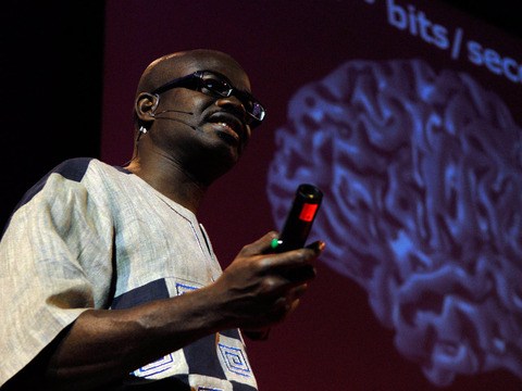
This is a thousand-year-old drawing of the brain. It's a diagram of the visual system. And some things look very familiar today. Two eyes at the bottom, optic nerve flowing out from the back. There's a very large nose that doesn't seem to be connected to anything in particular.
Dit is een 1000 jaar oude tekening van het brein. Het is een doorsnede van het visuele systeem. Sommige dingen zien er nog hetzelfde uit als vandaag. Twee ogen onderaan, optische zenuw komende van achteraan. Een zeer grote neus die schijnbaar niet verbonden is met iets in het bijzonder.
And if we compare this to more recent representations of the visual system, you'll see that things have gotten substantially more complicated over the intervening thousand years. And that's because today we can see what's inside of the brain, rather than just looking at its overall shape.
Als we dit vergelijken met meer recente voorstellingen van het visuele systeem, zie je dingen die substantieel gecompliceerder zijn geworden over de voorbije duizend jaren. Dat is omdat we vandaag in het brein kunnen kijken in plaats van enkel naar de vorm.
Imagine you wanted to understand how a computer works and all you could see was a keyboard, a mouse, a screen. You really would be kind of out of luck. You want to be able to open it up, crack it open, look at the wiring inside. And up until a little more than a century ago, nobody was able to do that with the brain. Nobody had had a glimpse of the brain's wiring.
Stel je voor dat je wil weten hoe een computer werkt en alles wat je kon zien was een toetsenbord, een muis en een scherm. Je zou geen geluk hebben. Je wil het open doen, ontleden, vanbinnen naar de bedrading kijken. Tot een eeuw geleden was niemand in staat om dat met het brein te doen. Niemand had nog een glimp opgevangen van de bedrading van het brein.
And that's because if you take a brain out of the skull and you cut a thin slice of it, put it under even a very powerful microscope, there's nothing there. It's gray, formless. There's no structure. It won't tell you anything.
Want als je een brein uit een schedel neemt en er dunne sneetjes afsnijdt en onder een krachtige microscoop steekt, zie je niets. Het is grijs en vormeloos. Er is geen structuur. Het zegt je niets.
And this all changed in the late 19th century. Suddenly, new chemical stains for brain tissue were developed and they gave us our first glimpses at brain wiring. The computer was cracked open.
Dit veranderde allemaal einde 19e eeuw. Plots werden nieuwe chemische stoffen voor breinweefsel ontwikkeld Ze gaven ons de eerste beelden van de bedrading van een brein. De computer was opengebroken.
So what really launched modern neuroscience was a stain called the Golgi stain. And it works in a very particular way. Instead of staining all of the cells inside of a tissue, it somehow only stains about one percent of them. It clears the forest, reveals the trees inside. If everything had been labeled, nothing would have been visible. So somehow it shows what's there.
De moderne neurowetenschappen werden eigenlijk gelanceerd door de golgikleuring. Het werkt op een heel gelijkaardige manier. In plaats van alle cellen in het weefsel te kleuren, kleurt het maar ongeveer één procent ervan. Het verheldert het bos, je ziet de bomen erin. Mocht alles gelabeld zijn, zou je niets meer zien. Op de een op andere manier toont het wat er is.
Spanish neuroanatomist Santiago Ramon y Cajal, who's widely considered the father of modern neuroscience, applied this Golgi stain, which yields data which looks like this, and really gave us the modern notion of the nerve cell, the neuron. And if you're thinking of the brain as a computer, this is the transistor. And very quickly Cajal realized that neurons don't operate alone, but rather make connections with others that form circuits just like in a computer. Today, a century later, when researchers want to visualize neurons, they light them up from the inside rather than darkening them. And there's several ways of doing this. But one of the most popular ones involves green fluorescent protein. Now green fluorescent protein, which oddly enough comes from a bioluminescent jellyfish, is very useful. Because if you can get the gene for green fluorescent protein and deliver it to a cell, that cell will glow green -- or any of the many variants now of green fluorescent protein, you get a cell to glow many different colors.
De Spaanse neuro-anatomist Santiage Ramon y Cajal, die als de vader van de moderne neurowetenschap wordt beschouwd, paste deze golgikleuring toe, en bekwam data die er zo uitzagen. Ze gaven ons de moderne visie op zenuwcellen, het neuron. Als je denkt aan een brein zoals aan een computer, dan is dit de transistor. Zeer snel realiseerde Cajal zich dat neuronen niet op zichzelf werken, maar verbinding maken met anderen die circuits vormen net zoals in een computer. Vandaag, een eeuw later, als onderzoekers neuronen willen voorstellen, verlichten ze deze in plaats van ze te verdonkeren. Er zijn verschillende manieren om dat te doen. Eén van de meest populaire gebeurt met groen fluorescerend eiwit. Groen fluorescerend eiwit, dat eigenaardig genoeg afkomstig is van een lichtgevende kwal, is zeer bruikbaar. Omdat als je het gen voor groen fluorescerend eiwit kan vinden, het afleveren aan een cel, zal die cel groen oplichten -- of een of andere variant van groen fluorescerend eiwit, krijg je een cel die in verschillende kleuren oplicht.
And so coming back to the brain, this is from a genetically engineered mouse called "Brainbow." And it's so called, of course, because all of these neurons are glowing different colors.
Ik keer terug naar het brein. Dit is een genetisch veranderde muis met de naam 'Brainbow'. Zo wordt ze genoemd, natuurlijk, omdat alle neuronen in een verschillende kleur oplichten.
Now sometimes neuroscientists need to identify individual molecular components of neurons, molecules, rather than the entire cell. And there's several ways of doing this, but one of the most popular ones involves using antibodies. And you're familiar, of course, with antibodies as the henchmen of the immune system. But it turns out that they're so useful to the immune system because they can recognize specific molecules, like, for example, the coat protein of a virus that's invading the body. And researchers have used this fact in order to recognize specific molecules inside of the brain, recognize specific substructures of the cell and identify them individually.
Soms moeten neurowetenschappers individuele moleculaire componenten van neuronen, moleculen, identificeren in plaats van een volledige cel. Er zijn verschillende manieren om dat te doen, maar in een van de meest populaire gebruiken ze antilichamen. Je bent natuurlijk wel bekend met antilichamen zoals de handlangers van het immuunsysteem. Het blijkt dat ze zo bruikbaar zijn voor het immuunsysteem omdat ze specifieke moleculen kunnen herkennen, zoals, bijvoorbeeld, de eiwitcode van een virus dat het lichaam binnendringt. Onderzoekers hebben dit feit gebruikt om specifieke moleculen in het brein te herkennen, specifieke substructuren van de cel te herkennen en ze individueel te identificeren.
And a lot of the images I've been showing you here are very beautiful, but they're also very powerful. They have great explanatory power. This, for example, is an antibody staining against serotonin transporters in a slice of mouse brain.
Een hoop van deze beelden die ik jullie getoond heb, zijn heel mooi maar ze zijn ook zeer krachtig. Ze hebben een grote verklarende kracht. Dit, bijvoorbeeld, is een antilichaamkleuring tegen serotonine-overbrengers in een schijfje van een muizenbrein.
And you've heard of serotonin, of course, in the context of diseases like depression and anxiety. You've heard of SSRIs, which are drugs that are used to treat these diseases. And in order to understand how serotonin works, it's critical to understand where the serontonin machinery is. And antibody stainings like this one can be used to understand that sort of question.
Je hebt natuurlijk al gehoord van serotonine in de context van ziektes zoals depressie en angst. Je heb gehoord van SSRI's, die medicijnen zijn die gebruikt worden om deze ziektes te genezen. Om te begrijpen hoe serotonine werkt, is het belangrijk om te verstaan waar deze serotonine-machinerie zit. Antilichaamkleuring zoals deze kan gebruikt worden om dat soort vragen te begrijpen.
I'd like to leave you with the following thought: Green fluorescent protein and antibodies are both totally natural products at the get-go. They were evolved by nature in order to get a jellyfish to glow green for whatever reason, or in order to detect the coat protein of an invading virus, for example. And only much later did scientists come onto the scene and say, "Hey, these are tools, these are functions that we could use in our own research tool palette." And instead of applying feeble human minds to designing these tools from scratch, there were these ready-made solutions right out there in nature developed and refined steadily for millions of years by the greatest engineer of all. Thank you. (Applause)
Ik wil jullie nog de volgende gedachte voorleggen: groen fluorescerend eiwit en antilichamen zijn beide van in het begin totaal natuurlijke producten. Ze zijn ontwikkeld door de natuur om een kwal groen te doen oplichten, om welke reden dan ook, of om bijvoorbeeld de eiwitcode van een binnendringend virus te detecteren. Pas later verschenen wetenschappers op het toneel en zeiden: 'Hé, dit zijn middelen, functies die we kunnen gebruiken als aanvulling op onze eigen onderzoeksmiddelen.' In plaats dat zwakke menselijke breinen dit moesten ontwerpen uit het niets, waren dit pasklare oplossingen recht uit de natuur, ontwikkeld en verfijnd over miljoenen jaren door de grootste ingenieur van allemaal. Dank je (Applaus)