This is a thousand-year-old drawing of the brain. It's a diagram of the visual system. And some things look very familiar today. Two eyes at the bottom, optic nerve flowing out from the back. There's a very large nose that doesn't seem to be connected to anything in particular.
이 그림은 천년전에 뇌의 모양을 그린 그림입니다. 이것은 시각계의 다이어그램 인데요. 오늘날 봐도 비슷한 부분이 많죠. 밑에 두개의 안구가 있고, 시신경이 뒤로부터 흐르고 있죠. 매우 큰 코도 있는데요. 특별히 다른 어떤 것과도 연결되 보이지 않네요.
And if we compare this to more recent representations of the visual system, you'll see that things have gotten substantially more complicated over the intervening thousand years. And that's because today we can see what's inside of the brain, rather than just looking at its overall shape.
우리가 이것을 최근에 만들어진 시각계의 그림과 비교하면, 여러분은 뇌 구조가 상당히 복잡해진 것을 알 수 있습니다. 그 사이 수천년 동안 말이죠. 그건 오늘날에 우리가 뇌의 내부를 볼 수 있기 때문입니다. 뇌의 그냥 전체적인 형태 말고도요.
Imagine you wanted to understand how a computer works and all you could see was a keyboard, a mouse, a screen. You really would be kind of out of luck. You want to be able to open it up, crack it open, look at the wiring inside. And up until a little more than a century ago, nobody was able to do that with the brain. Nobody had had a glimpse of the brain's wiring.
이제 여러분들이 컴퓨터의 작동원리를 알고 싶어한다고 상상해보죠, 그리고 볼 수 있는 것이라고는 키보드, 마우스, 스크린이라고 생각해봅시다. 그럼 정말 운이 없는거죠. 여러분은 컴퓨터를 열어 볼수있기를 바라고, 내부의 전선들을 보고 싶어 합니다. 한 세기 전까지만해도, 아무도 뇌를 그렇게 열어 볼 수 없었습니다. 아무도 뇌속의 신경들에 대해 알 수 없었습니다.
And that's because if you take a brain out of the skull and you cut a thin slice of it, put it under even a very powerful microscope, there's nothing there. It's gray, formless. There's no structure. It won't tell you anything.
왜냐하면 아무리 두개골 밖으로 뇌를 꺼내서 얇게 뇌를 자른 후, 고성능 현미경 밑에 올려봐도, 아무것도 안 보이기 때문이죠. 그냥 회색에, 형태가 없죠. 어떤 구조도 안가지고 있어 배울게 없다는 말입니다.
And this all changed in the late 19th century. Suddenly, new chemical stains for brain tissue were developed and they gave us our first glimpses at brain wiring. The computer was cracked open.
그러다가 19세기말에 모든것이 바뀌었습니다. 갑자기 뇌조직을 볼 수 있는 화학 착색제가 개발된것이죠. 그리고 뇌속의 신경들을 처음 볼 수 있게 해주었습니다. 컴퓨터 내부를 볼 수 있게 된거죠.
So what really launched modern neuroscience was a stain called the Golgi stain. And it works in a very particular way. Instead of staining all of the cells inside of a tissue, it somehow only stains about one percent of them. It clears the forest, reveals the trees inside. If everything had been labeled, nothing would have been visible. So somehow it shows what's there.
그래서 현대 신경과학을 부흥시킨 것은 골지(Golgi) 착색제라고 불리우는 재료입니다. 이 착색제는 특별한 방법으로 작용하게 되는데요. 조직내의 모든 세포에 착색하지 않고, 약 1%정도에만 착색하게 됩니다. 숲을 치워 버리고 그 안의 나무를 보이게 하는 것처럼 말이죠. 만약 모든 조직에 착색이 된다면, 아무것도 보지 못했을 겁니다. 그래서 이런 방법으로 조직내 신경들을 보여주기 시작한거죠.
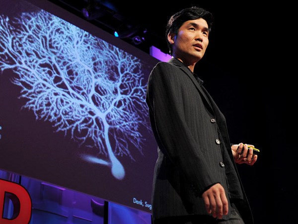
Spanish neuroanatomist Santiago Ramon y Cajal, who's widely considered the father of modern neuroscience, applied this Golgi stain, which yields data which looks like this, and really gave us the modern notion of the nerve cell, the neuron. And if you're thinking of the brain as a computer, this is the transistor. And very quickly Cajal realized that neurons don't operate alone, but rather make connections with others that form circuits just like in a computer. Today, a century later, when researchers want to visualize neurons, they light them up from the inside rather than darkening them. And there's several ways of doing this. But one of the most popular ones involves green fluorescent protein. Now green fluorescent protein, which oddly enough comes from a bioluminescent jellyfish, is very useful. Because if you can get the gene for green fluorescent protein and deliver it to a cell, that cell will glow green -- or any of the many variants now of green fluorescent protein, you get a cell to glow many different colors.
스페인의 신경해부학자 산티아고 라모니 카할은 현대 신경과학의 아버지로 불리우는데요, 골지 착색제를 이용해서 이렇게 보이는 자료를 만들어냈습니다. 그리고 신경세포와 뉴런에 대한 현대적인 개념을 정립했습니다. 그래서 여러분들이 뇌를 컴퓨터에 비유한다면, 이건 트랜지스터가 되는거죠. 그리고 카할은 뉴런들은 혼자서 작동하지 않는다는 것을 금새 알게 되었습니다. 대신에 컴퓨터안의 회로도 처럼 서로 연결되어 작동한다는것을 알게 된 것이죠. 한세기가 지난 오늘날, 연구원들이 뉴런을 보고자 하면, 뉴런들을 어둡게 하지 않고 밝게해서 보게 됩니다. 이런거에는 여러가지 방법이 있는데요. 많이 쓰는 방법중 하나는 녹색의 형광 단백질을 착색하는겁니다. 생체 발광 해파리로 부터 많이 얻을 수 있는 녹색 형광 단백질은 매우 유용합니다. 왜냐하면 녹색 형광 단백질에서 유전자를 채취해 세포에 주입하면, 그 세포는 녹색으로 발광하게 됩니다. -- 또는 녹색 형광 단백질의 다양한 변종으로 되거나요. 그래서 다양한 색깔로 빛나는 세포를 얻을 수 있기 때문에 유용합니다.
And so coming back to the brain, this is from a genetically engineered mouse called "Brainbow." And it's so called, of course, because all of these neurons are glowing different colors.
다시 뇌 이야기로 돌아와서요, 이 사진은 "Brainbow"라고 불리는 유전자 조작된 쥐에서 나온 것입니다. 이름에서 말하듯이 이 뉴런들이 서로 다른 색깔로 발광하게 됩니다.
Now sometimes neuroscientists need to identify individual molecular components of neurons, molecules, rather than the entire cell. And there's several ways of doing this, but one of the most popular ones involves using antibodies. And you're familiar, of course, with antibodies as the henchmen of the immune system. But it turns out that they're so useful to the immune system because they can recognize specific molecules, like, for example, the coat protein of a virus that's invading the body. And researchers have used this fact in order to recognize specific molecules inside of the brain, recognize specific substructures of the cell and identify them individually.
신경과학자들은 때로는 전체 세포보다는 뉴런의 각각의 분자 성분을 확인하고 싶을때가 있습니다. 몇가지 방법으로 확인 할 수 있는데요, 가장 보편적인 방법중에 하나는 항체를 이용하는 것입니다. 물론 면역체계에서 중요한 역할을 하는걸로 친숙하죠. 항체는 면역체계에 아주 유용한걸로 알려졌는데요, 특별한 분자를 인지 할 수 있기 때문입니다. 예를들어, 인체에 침투하는 바이러스의 단백질 코드 같은 것 말이죠. 그래서 연구원들이 뇌속의 특별한 분자를 찾아내기 위해 이런 사실을 이용하게 된 것입니다. 그리고 세포의 하부 구조들 각각을 구분하는데 이용하기도 합니다.
And a lot of the images I've been showing you here are very beautiful, but they're also very powerful. They have great explanatory power. This, for example, is an antibody staining against serotonin transporters in a slice of mouse brain.
오늘 여러분들에게 보여드린 많은 사진들은 정말 아름답기도 하지만, 매우 강력한 의미를 가지고 있습니다. 굉장히 설득력있는 이미지들입니다. 예들들어, 이것은 쥐의 뇌조직에서 세로틴에 반응하는 항체 착색 사진입니다.
And you've heard of serotonin, of course, in the context of diseases like depression and anxiety. You've heard of SSRIs, which are drugs that are used to treat these diseases. And in order to understand how serotonin works, it's critical to understand where the serontonin machinery is. And antibody stainings like this one can be used to understand that sort of question.
물론 여러분들은 우울증과 불안과 관련된 세로틴에 대해서 많이 들어 보셨을 겁니다. 이런 병들을 치료하는 우울증치료제(SSRI)도 들어 보셨을 겁니다. 세로틴이 어떻게 작용하는지 알기 위해서, 세로틴이 분비되는 곳이 어딘지를 아는것은 매우 중요합니다. 그래서 이와 같은 항체 착색은 이런 질문에 대한 답을 얻는데 이용 될 수 있습니다.
I'd like to leave you with the following thought: Green fluorescent protein and antibodies are both totally natural products at the get-go. They were evolved by nature in order to get a jellyfish to glow green for whatever reason, or in order to detect the coat protein of an invading virus, for example. And only much later did scientists come onto the scene and say, "Hey, these are tools, these are functions that we could use in our own research tool palette." And instead of applying feeble human minds to designing these tools from scratch, there were these ready-made solutions right out there in nature developed and refined steadily for millions of years by the greatest engineer of all. Thank you. (Applause)
저는 여러분들이 이런 생각들을 해주셨으면 합니다 : 녹색 형광 단백질과 항체 착색은 원래부터 천연 물질입니다. 그것들은 자연적으로 진화했는데, 뭐 예를 들면, 해파리가 초록색으로 보이게 하거나, 침투한 바이러스의 단백질 코드를 탐지하기 위해서 말입니다. 그리고 한참후에 과학자들이 나타나서 말하죠. "이야~ 여기 좋은 도구들이 있네!! 이 도구의 기능들은 우리의 연구에 사용해도 됬었을 텐데.." 보잘것 없는 인간의 생각을 활용하기 보다는 이런 도구들을 만들어 내기 위해, 위대한 기술자(자연)에 의해서 수백만년동안 잘 개발되고 다듬어진 자연적인 도구들이 존재한다는 것이죠. 감사합니다. (박수)