I will start by posing a little bit of a challenge: the challenge of dealing with data, data that we have to deal with in medical situations. It's really a huge challenge for us. And this is our beast of burden -- this is a Computer Tomography machine, a CT machine. It's a fantastic device. It uses X-rays, X-ray beams, that are rotating very fast around the human body. It takes about 30 seconds to go through the whole machine and is generating enormous amounts of information that comes out of the machine. So this is a fantastic machine that we can use for improving health care, but as I said, it's also a challenge for us. And the challenge is really found in this picture here. It's the medical data explosion that we're having right now. We're facing this problem. And let me step back in time.
Я начну с постановки довольно сложной задачи - задачи работы с данными, с теми данными, с которыми мы сталкиваемся в медицинском контексте. Это действительно очень сложная задача, над решением которой мы бьёмся день и ночь. Это компьютерный томограф - или просто КТ. Это фантастическое устройство. Оно использует рентгеновские лучи, которые очень быстро вращаются вокруг тела человека. Сканирование происходит за 30 секунд, и при этом томограф генерирует невероятный объём информации. Это потрясающая машина, с помощью которой мы можем улучшить здравоохранение. Но, как я уже сказал, это и сложная задача. На этом снимке можно увидеть эту задачу. Это настоящий бум медицинских данных, который мы наблюдаем в наше время. Мы активно ищем решение этой проблемы. Позвольте мне вернуться в недалёкое прошлое.
Let's go back a few years in time and see what happened back then. These machines that came out -- they started coming in the 1970s -- they would scan human bodies, and they would generate about 100 images of the human body. And I've taken the liberty, just for clarity, to translate that to data slices. That would correspond to about 50 megabytes of data, which is small when you think about the data we can handle today just on normal mobile devices. If you translate that to phone books, it's about one meter of phone books in the pile. Looking at what we're doing today with these machines that we have, we can, just in a few seconds, get 24,000 images out of a body, and that would correspond to about 20 gigabytes of data, or 800 phone books, and the pile would then be 200 meters of phone books. What's about to happen -- and we're seeing this; it's beginning -- a technology trend that's happening right now is that we're starting to look at time-resolved situations as well. So we're getting the dynamics out of the body as well. And just assume that we will be collecting data during five seconds, and that would correspond to one terabyte of data -- that's 800,000 books and 16 kilometers of phone books. That's one patient, one data set. And this is what we have to deal with.
Давайте вернёмся на несколько лет назад и посмотрим, что происходило тогда. Машины которые были разработаны - они начали появляться в 1970-е годы - сканировали организм человека и генерировали порядка 100 изображений человеческого организма. И я взял на себя смелость, просто для ясности, перевести это в объёмы данных. 100 снимков соответствуют примерно 50 Мб данных, что немного, если подумать об объёме данных, с которыми мы работаем сегодня на обычных мобильных устройствах. Если представить эти 100 изображений в виде телефонных книг, получится стопка телефонных справочников высотой 1 метр. Если мы вернёмся в сегодняшний день, с нынешними машинами мы можем всего за несколько секунд получить 24000 изображений организма. А это соответствует примерно 20 Гб данных или 800 телефонным книгам. И стопка телефонных справочников будет высотой 200 метров. То что произойдёт в скором времени - а мы уже видим начало этого процесса - при нынешних технологических тенденциях мы начинаем рассматривать ещё и временной фактор. Т.е. мы получаем ещё и информацию о динамике в организме. И если только представить, что мы будем собирать данные в течение 5 секунд, то это будет соответствовать терабайту данных. Это 800000 книг или 16 км телефонных справочников. И это один пациент, один набор данных. И с этим нам придётся справляться.
So this is really the enormous challenge that we have. And already today -- this is 25,000 images. Imagine the days when we had radiologists doing this. They would put up 25,000 images, they would go like this, "25,0000, okay, okay. There is the problem." They can't do that anymore. That's impossible. So we have to do something that's a little bit more intelligent than doing this. So what we do is that we put all these slices together. Imagine that you slice your body in all these directions, and then you try to put the slices back together again into a pile of data, into a block of data. So this is really what we're doing. So this gigabyte or terabyte of data, we're putting it into this block. But of course, the block of data just contains the amount of X-ray that's been absorbed in each point in the human body. So what we need to do is to figure out a way of looking at the things we do want to look at and make things transparent that we don't want to look at. So transforming the data set into something that looks like this. And this is a challenge. This is a huge challenge for us to do that.
Поэтому перед нами стоит действительно сложнейшая задача. И уже сегодня это 25000 изображений. Представьте себе время, когда рентгенологи анализировали снимки. Они бы повесили 25000 снимков и делали бы так: "25000, ну да, ну да. Вот, где проблема". Они уже больше не могут так работать; это невозможно. Поэтому мы должны придумать что-то поумней. И вот что мы делаем: мы складываем все снимки вместе. Представьте, что вы разрезаете организм во всех этих направлениях, а потом вы пытаетесь собрать кусочки вместе в стопку данных, в блок данных. Мы так и делаем. Мы берём гигабайт или терабайт данных и складываем их в блок. Этот блок данных содержит только то количество рентгеновских лучей, которое было поглощено каждой точкой тела. Поэтому нам надо найти способ посмотреть на то, на что мы хотим посмотреть, и сделать прозрачным то, что нам не нужно видеть. Поэтому мы преобразуем набор данных во что-то, что выглядит примерно так. А вот это - непростая задача. Это очень серьёзная задача.
Using computers, even though they're getting faster and better all the time, it's a challenge to deal with gigabytes of data, terabytes of data and extracting the relevant information. I want to look at the heart. I want to look at the blood vessels. I want to look at the liver. Maybe even find a tumor, in some cases. So this is where this little dear comes into play. This is my daughter. This is as of 9 a.m. this morning. She's playing a computer game. She's only two years old, and she's having a blast. So she's really the driving force behind the development of graphics-processing units. As long as kids are playing computer games, graphics is getting better and better and better. So please go back home, tell your kids to play more games, because that's what I need.
С помощью компьютеров, даже несмотря на их стремительное развитие, сложно иметь дело с гигабайтами данных, с терабайтами данных, и выуживать из них нужную информацию. Я хочу взглянуть на сердце, на кровеносные сосуды, на печень, может быть, даже обнаружить опухоль в определённых случаях. И вот здесь в игру вступает эта милашка. Это моя дочка. 9 утра. Она играет в компьютерную игру. Ей всего два годика, и ей ужасно весело. Поэтому она - настоящая движущая сила развития устройств для обработки графических изображений. Пока дети играют в компьютерные игры, графика будет становиться всё лучше и лучше. Поэтому, пожалуйста, скажите детям, пусть побольше играют в компьютерные игры, потому что мне это на руку.
So what's inside of this machine is what enables me to do the things that I'm doing with the medical data. So really what I'm doing is using these fantastic little devices. And you know, going back maybe 10 years in time when I got the funding to buy my first graphics computer -- it was a huge machine. It was cabinets of processors and storage and everything. I paid about one million dollars for that machine. That machine is, today, about as fast as my iPhone. So every month there are new graphics cards coming out, and here is a few of the latest ones from the vendors -- NVIDIA, ATI, Intel is out there as well. And you know, for a few hundred bucks you can get these things and put them into your computer, and you can do fantastic things with these graphics cards. So this is really what's enabling us to deal with the explosion of data in medicine, together with some really nifty work in terms of algorithms -- compressing data, extracting the relevant information that people are doing research on.
Внутренности этой машины позволяют мне обрабатывать медицинские данные. И моя работа состоит в использовании этих фантастических мелких устройств. Знаете, если мы вернёмся назад лет на 10, когда я получил грант на покупку первого графического компьютера, это была здоровая машина. Она занимала несколько комнат под процессоры, хранилища и т.д. Я заплатил около миллиона долларов за ту машину. Сейчас эта машина быстра, как мой iPhone. Каждый месяц выходят новые видеокарты Вот несколько новейших моделей - здесь есть NVIDIA, ATI, Intel. И теперь эти видеокарты можно купить за несколько сотен баксов, установить на компьютер. А с такими видеокартами можно делать потрясающие вещи. Они помогают нам справляться с бумом медицинских данных, а также с по-настоящему тонкой работой, с точки зрения алгоритмов: сжатие данных, извлечение нужной информации, на основе которой проводится исследование.
So I'm going to show you a few examples of what we can do. This is a data set that was captured using a CT scanner. You can see that this is a full data [set]. It's a woman. You can see the hair. You can see the individual structures of the woman. You can see that there is [a] scattering of X-rays on the teeth, the metal in the teeth. That's where those artifacts are coming from. But fully interactively on standard graphics cards on a normal computer, I can just put in a clip plane. And of course all the data is inside, so I can start rotating, I can look at it from different angles, and I can see that this woman had a problem. She had a bleeding up in the brain, and that's been fixed with a little stent, a metal clamp that's tightening up the vessel. And just by changing the functions, then I can decide what's going to be transparent and what's going to be visible. I can look at the skull structure, and I can see that, okay, this is where they opened up the skull on this woman, and that's where they went in. So these are fantastic images. They're really high resolution, and they're really showing us what we can do with standard graphics cards today.
Я хочу показать вам несколько примеров того, что теперь возможно. Это набор данных, снятых при помощи КТ-сканера. Вы видите, что он полон данных. Это женщина. Вы можете видеть волосы. Вы видите отдельные части тела женщины. Вы видите, как разбросаны рентгеновские лучи - на зубах, на металле в зубах. Вот откуда здесь помехи. Это происходит совершенно интерактивно, на стандартной видеокарте на обычном компьютере. Я могу просто вставить плоскость отсечения. И, конечно, все эти данные находятся внутри, поэтому я могу начать вращение, я могу посмотреть под любым углом, и я вижу, что у этой женщины были проблемы. У неё было кровотечение в мозгу, и его вылечили с помощью стента, металлической скобки, которая пережала сосуд. Просто поменяв функции, я могу решить, что должно быть прозрачным, а что должно быть видимым. Я могу взглянуть на структуру черепа, и я вижу, что вот здесь череп был вскрыт, а здесь произошло проникновение внутрь. Это потрясающие изображения. Очень высокого разрешения, и они показывают нам, что мы можем сделать с обычными видеокартами уже сегодня.
Now we have really made use of this, and we have tried to squeeze a lot of data into the system. And one of the applications that we've been working on -- and this has gotten a little bit of traction worldwide -- is the application of virtual autopsies. So again, looking at very, very large data sets, and you saw those full-body scans that we can do. We're just pushing the body through the whole CT scanner, and just in a few seconds we can get a full-body data set. So this is from a virtual autopsy. And you can see how I'm gradually peeling off. First you saw the body bag that the body came in, then I'm peeling off the skin -- you can see the muscles -- and eventually you can see the bone structure of this woman.
Итак, мы воспользовались видеокартой и попробовали впихнуть кучу данных в систему. Одно из приложений, над которым мы работали оно уже начинает использоваться по всему миру - это приложение виртуальной аутопсии (вскрытия). И снова, глядя на огромные наборы данных, а вы видели, какие виды сканирования всего тела мы можем делать. Мы проводим тело через компьютерный томограф, и через несколько секунд получаем полный набор данных. Виртуальная аутопсия позволяет получить эту информацию. Вы видите, как я постепенно снимаю слой за слоем. Сначала вы видите мешок для перевозки трупа, в котором поступило тело, затем я снимаю кожу - вы видите мускулы, и, наконец, вы видите костную структуру этой женщины.
Now at this point, I would also like to emphasize that, with the greatest respect for the people that I'm now going to show -- I'm going to show you a few cases of virtual autopsies -- so it's with great respect for the people that have died under violent circumstances that I'm showing these pictures to you. In the forensic case -- and this is something that ... there's been approximately 400 cases so far just in the part of Sweden that I come from that has been undergoing virtual autopsies in the past four years. So this will be the typical workflow situation. The police will decide -- in the evening, when there's a case coming in -- they will decide, okay, is this a case where we need to do an autopsy? So in the morning, in between six and seven in the morning, the body is then transported inside of the body bag to our center and is being scanned through one of the CT scanners. And then the radiologist, together with the pathologist and sometimes the forensic scientist, looks at the data that's coming out, and they have a joint session. And then they decide what to do in the real physical autopsy after that.
Здесь я бы хотел подчеркнуть, что испытываю огромное уважение к людям, которых я сейчас покажу - я собираюсь показать несколько виртуальных аутопсий, я делаю это с огромным уважением к людям, которые погибли при страшных обстоятельствах, и чьи снимки я покажу вам. В судебной медицине до сих пор у нас было около 400 таких случаев только в той части Швеции, откуда я родом, в последние 4 года в таких случаях проводится виртуальная аутопсия. Поэтому это типичный рабочий процесс. Полиция принимает решение - вечером, когда произошло происшествие, они решают, что в этом случае им понадобится аутопсия. И утром, между 6 и 7 часами, тело привозят в мешке для перевозки трупов в наш центр, его сканируют при помощи компьютерного томографа. И потом рентгенолог вместе с патологоанатомом и иногда судебным экспертом смотрят на полученные данные и проводят консилиум. И после этого они решают, что нужно сделать во время реальной аутопсии.
Now looking at a few cases, here's one of the first cases that we had. You can really see the details of the data set. It's very high-resolution, and it's our algorithms that allow us to zoom in on all the details. And again, it's fully interactive, so you can rotate and you can look at things in real time on these systems here. Without saying too much about this case, this is a traffic accident, a drunk driver hit a woman. And it's very, very easy to see the damages on the bone structure. And the cause of death is the broken neck. And this women also ended up under the car, so she's quite badly beaten up by this injury.
Посмотрим на несколько случаев: это один из первых. Вы видите подробности набора данных; это очень высокое разрешение. И наши алгоритмы позволяют нам приблизить любую деталь. Повторю, это полностью интерактивный процесс, поэтому на этих системах изображение можно повернуть и посмотреть в реальном времени. Не говоря лишних слов о происшествии, это было ДТП, пьяный водитель сбил женщину. И здесь очень просто увидеть повреждения костной структуры. Причиной смерти стал перелом шеи. Женщина оказалась под машиной, поэтому на теле довольно много повреждений.
Here's another case, a knifing. And this is also again showing us what we can do. It's very easy to look at metal artifacts that we can show inside of the body. You can also see some of the artifacts from the teeth -- that's actually the filling of the teeth -- but because I've set the functions to show me metal and make everything else transparent. Here's another violent case. This really didn't kill the person. The person was killed by stabs in the heart, but they just deposited the knife by putting it through one of the eyeballs. Here's another case. It's very interesting for us to be able to look at things like knife stabbings. Here you can see that knife went through the heart. It's very easy to see how air has been leaking from one part to another part, which is difficult to do in a normal, standard, physical autopsy. So it really, really helps the criminal investigation to establish the cause of death, and in some cases also directing the investigation in the right direction to find out who the killer really was.
Вот другой случай, поножовщина. Он тоже показателен, с точки зрения того, что мы можем сделать. Очень просто рассматривать металлические предметы, которые мы можем показать внутри тела. Вы видите помехи из-за зубов - это зубные пломбы - поскольку я настроил функции на показ металла, а всё остальное сделал прозрачным. Это другой жуткий случай. Не это убило этого человека. Человек был убит ударами ножом в сердце, но убийцы оставили нож, воткнув его в одно из глазных яблок. Это другой случай. Очень интересный для нас, поскольку мы можем разглядеть удары ножом. Здесь вы видите, что нож прошёл через сердце. Хорошо видно, как воздух перетекал из одной части в другую, это очень сложно увидеть в нормальной, стандартной, физической аутопсии. Поэтому виртуальная аутопсия по-настоящему помогает уголовным расследованиям установить причину смерти, а в некоторых случаях также задаёт следствию нужное направление для установления настоящего убийцы.
Here's another case that I think is interesting. Here you can see a bullet that has lodged just next to the spine on this person. And what we've done is that we've turned the bullet into a light source, so that bullet is actually shining, and it makes it really easy to find these fragments. During a physical autopsy, if you actually have to dig through the body to find these fragments, that's actually quite hard to do.
Это ещё один, на мой взгляд, интересный случай. Здесь вы видите пулю которая оказалась рядом с позвоночником. Что мы сделали? Мы превратили пулю в источник света, так что она светилась, и нам было легко обнаружить эти фрагменты. Во время физической аутопсии, приходится копаться в теле, чтобы найти эти фрагменты, это довольно сложно сделать.
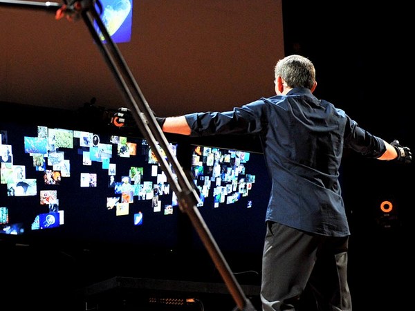
One of the things that I'm really, really happy to be able to show you here today is our virtual autopsy table. It's a touch device that we have developed based on these algorithms, using standard graphics GPUs. It actually looks like this, just to give you a feeling for what it looks like. It really just works like a huge iPhone. So we've implemented all the gestures you can do on the table, and you can think of it as an enormous touch interface. So if you were thinking of buying an iPad, forget about it. This is what you want instead. Steve, I hope you're listening to this, all right. So it's a very nice little device. So if you have the opportunity, please try it out. It's really a hands-on experience. So it gained some traction, and we're trying to roll this out and trying to use it for educational purposes, but also, perhaps in the future, in a more clinical situation. There's a YouTube video that you can download and look at this, if you want to convey the information to other people about virtual autopsies.
Предмет моей особенной гордости, который я счастлив представить вам сегодня, - это наш стол для виртуальной аутопсии. Это сенсорное устройство, которое мы разработали на основе этих алгоритмов на стандартном графическом процессоре. Вот как он выглядит. просто чтобы дать вам представление о том, как он выглядит. Он работает, как огромный iPhone. И мы сумели смоделировать все жесты, которые можно сделать на столе, и получился как бы огромный сенсорный интерфейс. Если вы подумываете, не купить ли вам iPad, забудьте о нём; вот, что вам нужно на самом деле. Стив, я надеюсь, ты это слышишь. Итак, это милое крошечное устройство. Если у вас есть возможность, пожалуйста, испытайте его. Здесь важен практический опыт. Этот проект набирает ход, мы надеемся его раскрутить и использовать в образовательных целях, а тажке, возможно, в будущем чаще применять его в клинических условиях. Вот ролик на YouTube, который вы можете скачать и посмотреть, если вы хотите рассказать другим людям о виртуальной аутопсии.
Okay, now that we're talking about touch, let me move on to really "touching" data. And this is a bit of science fiction now, so we're moving into really the future. This is not really what the medical doctors are using right now, but I hope they will in the future. So what you're seeing on the left is a touch device. It's a little mechanical pen that has very, very fast step motors inside of the pen. And so I can generate a force feedback. So when I virtually touch data, it will generate forces in the pen, so I get a feedback. So in this particular situation, it's a scan of a living person. I have this pen, and I look at the data, and I move the pen towards the head, and all of a sudden I feel resistance. So I can feel the skin. If I push a little bit harder, I'll go through the skin, and I can feel the bone structure inside. If I push even harder, I'll go through the bone structure, especially close to the ear where the bone is very soft. And then I can feel the brain inside, and this will be the slushy like this.
Раз уж мы заговорили об устройствах, работающих от прикосновения, давайте перейдём к данным, получаемым с помощью этих устройств. Сейчас начнётся научная фантастика, и мы переместимся в будущее. Врачи пока используют не совсем такое оборудование, но, я надеюсь, в будущем ситуация изменится. Слева вы видите сенсорное устройство. Это маленькая механическая ручка, внутри которой находятся исключительно быстрые шаговые регуляторы. И я могу сгенерировать силовую обратную связь. Поэтому когда я виртуально касаюсь данных, я генерирую силу прикосновения в ручке и получаю обратную связь. В этом случае мы видим результат сканирования живого человека. У меня есть ручка, я смотрю на данные, и я двигаю ручку к голове, и вдруг я чувствую сопротивление. И я могу пощупать кожу. Если я надавлю чуть сильнее, я пройду через кожу и смогу увидеть костную структуру изнутри. Если я надавлю ещё сильнее, я пройду сквозь костную структуру, особенно рядом с ухом, где кость очень мягкая. И тогда я увижу мозг внутри, он выглядит, как слякоть.
So this is really nice. And to take that even further, this is a heart. And this is also due to these fantastic new scanners, that just in 0.3 seconds, I can scan the whole heart, and I can do that with time resolution. So just looking at this heart, I can play back a video here. And this is Karljohan, one of my graduate students who's been working on this project. And he's sitting there in front of the Haptic device, the force feedback system, and he's moving his pen towards the heart, and the heart is now beating in front of him, so he can see how the heart is beating. He's taken the pen, and he's moving it towards the heart, and he's putting it on the heart, and then he feels the heartbeats from the real living patient. Then he can examine how the heart is moving. He can go inside, push inside of the heart, and really feel how the valves are moving. And this, I think, is really the future for heart surgeons. I mean it's probably the wet dream for a heart surgeon to be able to go inside of the patient's heart before you actually do surgery, and do that with high-quality resolution data. So this is really neat.
И это очень здорово. Пойдёмте дальше, вот сердце. Снова благодаря этим фантастическим сканнерам всего за 0,3 секунды я могу просканировать всё сердце, более того, я могу сделать это с временным разрешением. Глядя на это сердце, Вот здесь я могу снова просмотреть видео. А это Карл-Йохан, один из моих аспирантов, который работает над этим проектом. Он сидит за тактильным устройством, системой силовой обратной связи, и ведёт ручку к сердцу, и теперь сердце бьётся перед ним, и он может наблюдать, как бьётся сердце. Он берёт ручку и двигает её к сердцу, ставит её на сердце и чувствует сердцебиение реального живого человека. Затем он может исследовать движения сердца. Он может зайти внутрь, толкнуть сердце изнутри и почувствовать, как двигаются клапаны. И я думаю, что это и есть будущее кардиохирургов. Я имею в виду, что кардиохирурги, возможно, только мечтают о том, чтобы иметь возможность зайти внутрь сердца пациента до начала настоящей операции, и сделать это на основе данных с разрешением высокого качества. И это замечательно.
Now we're going even further into science fiction. And we heard a little bit about functional MRI. Now this is really an interesting project. MRI is using magnetic fields and radio frequencies to scan the brain, or any part of the body. So what we're really getting out of this is information of the structure of the brain, but we can also measure the difference in magnetic properties of blood that's oxygenated and blood that's depleted of oxygen. That means that it's possible to map out the activity of the brain. So this is something that we've been working on. And you just saw Motts the research engineer, there, going into the MRI system, and he was wearing goggles. So he could actually see things in the goggles. So I could present things to him while he's in the scanner. And this is a little bit freaky, because what Motts is seeing is actually this. He's seeing his own brain. So Motts is doing something here, and probably he is going like this with his right hand, because the left side is activated on the motor cortex. And then he can see that at the same time. These visualizations are brand new. And this is something that we've been researching for a little while.
Теперь мы ещё глубже погрузимся в научную фантастику. Все вы слышали о функциональной магнитно-резонансной томографии (МРТ). Это по-настоящему интересный проект. МРТ использует магнитные поля и радиочастоты для сканирования мозга или других частей тела. В результате мы получаем данные о структуре мозга, а также мы можем измерить разницу магнитных свойств крови, насыщенной кислородом, и крови, ненасыщенной кислородом. Это означает, что возможно составить карту активности мозга. Вот над этим мы и работаем. Только что вы видели Моттса, инженера-исследователя, который проходит МРТ в дисплейных очках. Он мог видеть в этих дисплейных очках. Поэтому я мог показывать ему кое-что, пока он был внутри сканера. Это довольно удивительно, потому что Моттс видит вот это: он видит свой собственный мозг. Моттс что-то делает здесь. И, возможно, он собирается показать вот такой жест правой рукой, потому что слева активирована двигательная область коры головного мозга. И в то же время он всё это видит. Такие визуализации - это новая разработка. Мы проводим исследования в этой области.
This is another sequence of Motts' brain. And here we asked Motts to calculate backwards from 100. So he's going "100, 97, 94." And then he's going backwards. And you can see how the little math processor is working up here in his brain and is lighting up the whole brain. Well this is fantastic. We can do this in real time. We can investigate things. We can tell him to do things. You can also see that his visual cortex is activated in the back of the head, because that's where he's seeing, he's seeing his own brain. And he's also hearing our instructions when we tell him to do things. The signal is really deep inside of the brain as well, and it's shining through, because all of the data is inside this volume. And in just a second here you will see -- okay, here. Motts, now move your left foot. So he's going like this. For 20 seconds he's going like that, and all of a sudden it lights up up here. So we've got motor cortex activation up there. So this is really, really nice, and I think this is a great tool. And connecting also with the previous talk here, this is something that we could use as a tool to really understand how the neurons are working, how the brain is working, and we can do this with very, very high visual quality and very fast resolution.
Это ещё одна сессия исследования мозга Моттса. Здесь мы попросили его считать обратно со 100. И он начал: "100, 97, 94" И считал назад. И вы видите, как маленький математический процессор работает вот здесь в мозгу и озаряет весь мозг. Это потрясающе. Мы можем проделать это в реальном времени. Мы можем исследовать. Мы можем говорить ему, что делать. Вы видите, что в задней части головы активирована зрительная кора головного мозга, потому что ею он видит, видит свой собственный мозг. И он слышит наши инструкции, когда мы говорим ему, что делать. Сигнал проходит вглубь мозга, но он просвечивается, потому что все данные находятся внутри этого объёма. И через секунду вы увидите - вот, здесь. Моттс, пошевели левой ногой. И он делает так. 20 секунд он делал так, а потом вдруг здесь озарение. Произошла активация двигательной зоны коры головного мозга вон там. Это очень, очень интересно. И я думаю, что это прекрасный инструмент. И, в связи с предыдущей речью TEDtalk, это технология, которую мы можем использовать как инструмент, помогающий по-настоящему понять, как работают нейроны, как работает мозг, и мы можем работать с очень, очень высоким качеством изображения и очень быстрым разрешением.
Now we're also having a bit of fun at the center. So this is a CAT scan -- Computer Aided Tomography. So this is a lion from the local zoo outside of Norrkoping in Kolmarden, Elsa. So she came to the center, and they sedated her and then put her straight into the scanner. And then, of course, I get the whole data set from the lion. And I can do very nice images like this. I can peel off the layer of the lion. I can look inside of it. And we've been experimenting with this. And I think this is a great application for the future of this technology, because there's very little known about the animal anatomy. What's known out there for veterinarians is kind of basic information. We can scan all sorts of things, all sorts of animals. The only problem is to fit it into the machine. So here's a bear. It was kind of hard to get it in. And the bear is a cuddly, friendly animal. And here it is. Here is the nose of the bear. And you might want to cuddle this one, until you change the functions and look at this. So be aware of the bear.
Ну, а ещё у себя в центре мы любим повеселиться. Это КТ - кошачий томограф. А это львица Эльза из местного зоопарка который находится недалеко от города Норркопинг в Кольмордене. Её привезли в центр и дали ей успокоительное, а потом поместили прямо в сканер. А потом, конечно, я собрал все данные по льву. И теперь я могу распечатывать такие классные фотографии. И могу снять шкуру с неубитого льва. Я могу заглянуть внутрь животного. Мы экспериментировали с этими данными. Я думаю, у этой технологии большое практическое будущее. Потому что анатомия животных очень плохо изучена. То что знают ветеринары - это базовая информация. Мы можем сканировать что угодно, у любых животных. Единственная проблема - впихнуть животное в томограф. Вот медведь. Было немного сложно его туда впихнуть. Медведь - ласковое, дружелюбное создание. А вот и он. Вот медвежий нос. У вас возникнет желание его обнять, пока вы не поменяете функции и не взглянете вот на это. Опасайтесь медведей.
So with that, I'd like to thank all the people who have helped me to generate these images. It's a huge effort that goes into doing this, gathering the data and developing the algorithms, writing all the software. So, some very talented people. My motto is always, I only hire people that are smarter than I am and most of these are smarter than I am.
И на этом я бы хотел поблагодарить всех, кто помогал мне сгенерировать эти изображения. Это огромный труд по сбору данных и разработке алгоритмов и написанию программ. Это очень талантливые люди. Мой девиз - нанимать только тех, кто умнее меня, и большинство из них умней меня.
So thank you very much.
Большое спасибо.
(Applause)
(аплодисменты)