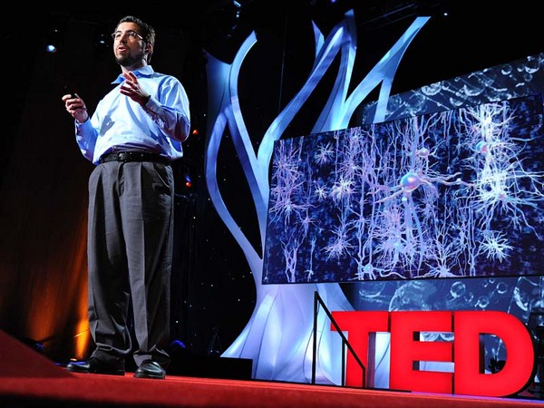
Humans have long held a fascination for the human brain. We chart it, we've described it, we've drawn it, we've mapped it. Now just like the physical maps of our world that have been highly influenced by technology -- think Google Maps, think GPS -- the same thing is happening for brain mapping through transformation.
Люди вже довгий час зачаровані людським мозком. Ми засхемували його, описали, ми зобразили його і нанесли на карту. Тепер, подібно до того, як на фізичні мапи світу неабиякий вплив здійснили технології – згадайте Google Maps, згадайте GPS – те саме відбувається і з мапуванням мозку, через трансформацію.
So let's take a look at the brain. Most people, when they first look at a fresh human brain, they say, "It doesn't look what you're typically looking at when someone shows you a brain." Typically, what you're looking at is a fixed brain. It's gray. And this outer layer, this is the vasculature, which is incredible, around a human brain. This is the blood vessels. 20 percent of the oxygen coming from your lungs, 20 percent of the blood pumped from your heart, is servicing this one organ. That's basically, if you hold two fists together, it's just slightly larger than the two fists.
Тож погляньмо на мозок. Більшість людей, що вперше дивляться на свіжий людський мозок, кажуть: "Він не схожий на те, на що ви зазвичай дивитесь, коли хтось показує вам мозок." Зазвичай ви дивитесь на затверділий мозок. Він сірий. І цей верхній шар – це судинна сітка, розташована довкола людського мозку. Це – кровоносні судини. 20% кисню, що надходить з ваших легенів, 20% крові, що відкачується з серця, обслуговують один цей орган. Фактично, якщо стиснути разом два кулаки, він трохи більший за ці два кулаки.
Scientists, sort of at the end of the 20th century, learned that they could track blood flow to map non-invasively where activity was going on in the human brain. So for example, they can see in the back part of the brain, which is just turning around there. There's the cerebellum; that's keeping you upright right now. It's keeping me standing. It's involved in coordinated movement. On the side here, this is temporal cortex. This is the area where primary auditory processing -- so you're hearing my words, you're sending it up into higher language processing centers. Towards the front of the brain is the place in which all of the more complex thought, decision making -- it's the last to mature in late adulthood. This is where all your decision-making processes are going on. It's the place where you're deciding right now you probably aren't going to order the steak for dinner.
Десь наприкінці 20-го сторіччя науковці з'ясували, що можуть відслідкувати шлях потоку крові, щоб без хірургічного втручання відобразити, де у людському мозку відбувається активність. Так, наприклад, вони можуть зазирнути у задню частину мозку, яка щойно була повернута до нас. Це мозочок; він відповідає за утримання вас у вертикальному положенні. Він дозволяє мені стояти. Він залучений у координування рухів. А з цього боку – скронева кора. Це область, де відбувається первинна обробка звуку – таким чином ви чуєте мої слова і відсилаєте їх далі, до високорівневих центрів обробки мови. Близько до фронтальної частини мозку знаходиться зона, відповідальна за комплексне мислення, прийняття рішень – вона формується останньою, вже в зрілому віці. Ось тут відбувається процес прийняття усіх ваших рішень. Це місце, де саме зараз ви вирішуєте, що стейк сьогодні на вечерю ви, мабуть, не замовите.
So if you take a deeper look at the brain, one of the things, if you look at it in cross-section, what you can see is that you can't really see a whole lot of structure there. But there's actually a lot of structure there. It's cells and it's wires all wired together. So about a hundred years ago, some scientists invented a stain that would stain cells. And that's shown here in the the very light blue. You can see areas where neuronal cell bodies are being stained. And what you can see is it's very non-uniform. You see a lot more structure there. So the outer part of that brain is the neocortex. It's one continuous processing unit, if you will. But you can also see things underneath there as well. And all of these blank areas are the areas in which the wires are running through. They're probably less cell dense. So there's about 86 billion neurons in our brain. And as you can see, they're very non-uniformly distributed. And how they're distributed really contributes to their underlying function. And of course, as I mentioned before, since we can now start to map brain function, we can start to tie these into the individual cells.
Якщо роздивлятися мозок більш детально, то одна з речей, яку можна побачити, дивлячись на нього у поперечному перерізі, це те, що тут відсутня певна чітка структура. Але насправді, структура там досить чітка. Всі клітини і канали мозку пов'язані. Тож близько ста років тому вчені винайшли барвник, здатний надати клітинам колір. І тут він показаний дуже світлим блакитним кольором. Ви можете бачити зони, де тіла нейронних клітин забарвлені. Як помітно, забарвлення дуже неоднорідне. Тут видно ще більшу структурованість. Далі, зовнішня частина людського мозку - це його кора. Це, так би мовити, єдиний сектор обробки даних. Але видно і речі, розташовані глибше під ним. Усі ці порожні ділянки - це зони, через які проходять сполучні волокна. Вони мають меншу густину клітин. У мозку людини близько 86 мільярдів нейронів. І, як видно, розподілені вони досить неоднорідно. Але саме такий розподіл сприяє їх основній функції. І звичайно, як я вже зазначав, оскільки ми вже можемо починати складати мапу для функцій мозку, ми можемо почати прив'язувати їх навіть до окремих клітин.
So let's take a deeper look. Let's look at neurons. So as I mentioned, there are 86 billion neurons. There are also these smaller cells as you'll see. These are support cells -- astrocytes glia. And the nerves themselves are the ones who are receiving input. They're storing it, they're processing it. Each neuron is connected via synapses to up to 10,000 other neurons in your brain. And each neuron itself is largely unique. The unique character of both individual neurons and neurons within a collection of the brain are driven by fundamental properties of their underlying biochemistry. These are proteins. They're proteins that are controlling things like ion channel movement. They're controlling who nervous system cells partner up with. And they're controlling basically everything that the nervous system has to do.
Давайте поглянемо більш детально. Розглянемо нейрони. Як я вже казав, нейронів близько 86 мільярдів. Додатково існують ще менші клітини, як видно тут. Це клітини підтримки - астрогліальні клітини. А самі нерви безпосередньо отримують вхідні дані. Вони зберігають та обробляють їх. Кожний нейрон пов'язаний за допомогою синапсів з понад 10 000 інших нейронів у вашому мозку. Та кожен нейрон майже абсолютно унікальний. Унікальність окремих нейронів та нейронів, сполучених при формуванні мозку, пояснюється фундаментальними властивостями їх базових біохімічних структур. А саме, протеїнів. Це білки, що контролюють такі процеси, як пересування по іонних каналах. Вони контролюють, як контактують клітини нервової системи. Фактично, вони керують усім, що має робити нервова система.
So if we zoom in to an even deeper level, all of those proteins are encoded by our genomes. We each have 23 pairs of chromosomes. We get one from mom, one from dad. And on these chromosomes are roughly 25,000 genes. They're encoded in the DNA. And the nature of a given cell driving its underlying biochemistry is dictated by which of these 25,000 genes are turned on and at what level they're turned on.
А якщо ми роздивимося більш детально, то виявиться, що всі протеїни закодовані нашими геномами. В кожного з нас 23 пари хромосомів. Ми беремо одну від матері, іншу від батька. І на цих хромосомах близько 25 000 генів. Вони закодовані в ДНК. Та сутність кожної даної клітини, що визначає її базову біохімію, продиктована тим, які саме з цих 25 000 генів активно залучені у роботу, та на якому рівні вони активовані.
And so our project is seeking to look at this readout, understanding which of these 25,000 genes is turned on. So in order to undertake such a project, we obviously need brains. So we sent our lab technician out. We were seeking normal human brains. What we actually start with is a medical examiner's office. This a place where the dead are brought in. We are seeking normal human brains. There's a lot of criteria by which we're selecting these brains. We want to make sure that we have normal humans between the ages of 20 to 60, they died a somewhat natural death with no injury to the brain, no history of psychiatric disease, no drugs on board -- we do a toxicology workup. And we're very careful about the brains that we do take. We're also selecting for brains in which we can get the tissue, we can get consent to take the tissue within 24 hours of time of death. Because what we're trying to measure, the RNA -- which is the readout from our genes -- is very labile, and so we have to move very quickly.
Таким чином, наш проект націлений на визначення та розуміння, які саме гени з цих 25 000 активовані. Для того, щоб братися до роботи над таким проектом, нам, звичайно, знадобиться мозок. Тож ми відправили нашого лаборанта на пошуки. Ми шукали нормальний людський мозок. Нашу роботу ми звичайно почали з офісу паталогоанатома. Це місце, куди привозять мерців. Ми шукаємо нормальний людський мозок. Для відбору мозку в нас є багато критеріїв. Ми хочемо впевнитись, що в нас нормальні люди, віком від 20 до 60 років, які померли від причин, близьких до природніх, не завдавши шкоди мозку, в яких не було психічних захворювань; які не приймали наркотичних речовин - для цього ми робимо токсикологічні аналізи. І ми дуже обережні з тими зразками, які обираємо наприкінці. Ми також маємо відібрати саме ті мізки, з яких ми можемо забрати тканину, тобто можемо отримати дозвіл взяти таку тканину не пізніше 24 годин з часу смерті. Тому що інформація, яку ми намагаємося виміряти, а саме РНК - її також можна назвати сумою показників, знятих з наших генів - є дуже нестійкою, тож ми мусимо працювати дуже швидко.
One side note on the collection of brains: because of the way that we collect, and because we require consent, we actually have a lot more male brains than female brains. Males are much more likely to die an accidental death in the prime of their life. And men are much more likely to have their significant other, spouse, give consent than the other way around.
Одна ремарка щодо обрання мозку: через спосіб, яким ми користуємося під час відбору, а також через те, що нам потрібна згода, в нас є набагато більше чоловічих мозків, ніж жіночих. Чоловіки набагато схильніші до випадкової смерті у розквіті життя. І набагато частіше їх друга половинка, дружина, дає згоду на донацію, ніж навпаки.
(Laughter)
(Сміх)
So the first thing that we do at the site of collection is we collect what's called an MR. This is magnetic resonance imaging -- MRI. It's a standard template by which we're going to hang the rest of this data. So we collect this MR. And you can think of this as our satellite view for our map. The next thing we do is we collect what's called a diffusion tensor imaging. This maps the large cabling in the brain. And again, you can think of this as almost mapping our interstate highways, if you will. The brain is removed from the skull, and then it's sliced into one-centimeter slices. And those are frozen solid, and they're shipped to Seattle. And in Seattle, we take these -- this is a whole human hemisphere -- and we put them into what's basically a glorified meat slicer. There's a blade here that's going to cut across a section of the tissue and transfer it to a microscope slide. We're going to then apply one of those stains to it, and we scan it. And then what we get is our first mapping.
Перше, що ми робимо на місці відбору, зберігаємо так званий МР. Це магніторезонансна томографія - МРТ. Це стандартний шаблон, до якого ми будемо додавати наступні дані. Тож ми зберігаємо Магнітний Резонанс. Його ще можна назвати супутниковим видом нашої мапи. Наступним кроком ми робимо візуалізацію мозку дифузно-тензорним методом. Це зображає схему нервових зв'язків. Знову ж таки, можна уявити це майже як нанесення на мапу головних швидкісних автомагістралей. Потім мозок виймається з черепа і нарізається шарами завширшки один сантиметр. Ці шари фіксуються за допомогою заморожування і відправляються до Сіетлу. А в Сіетлі ми беремо ось це - таку цілу півкулю людського мозку - і кладемо її у ніщо інше, як у славетну машину для нарізки м'яса. Її лезо відрізає секцію тканини і переносить її на слайд для мікроскопа. Потім ми застосуємо до нього один з тих барвників і зможемо його відсканувати. Таким чином ми отримаємо нашу першу мапу.
So this is where experts come in and they make basic anatomic assignments. You could consider this state boundaries, if you will, those pretty broad outlines. From this, we're able to then fragment that brain into further pieces, which then we can put on a smaller cryostat. And this is just showing this here -- this frozen tissue, and it's being cut. This is 20 microns thin, so this is about a baby hair's width. And remember, it's frozen. And so you can see here, old-fashioned technology of the paintbrush being applied. We take a microscope slide. Then we very carefully melt onto the slide. This will then go onto a robot that's going to apply one of those stains to it. And our anatomists are going to go in and take a deeper look at this.
А потім з'являться експерти, щоб зробити базове анатомічне обстеження. Про це можна думати як про нанесення регіональних меж, таке собі охайне межування. Після цього ми можемо розділяти мозок на сегменти, які потім помістяться у менший кріостат. Саме тут ви можете бачити якраз це - заморожену тканину і те, як вона розрізається. Цей сегмент завтовшки 20 мікронів, тобто як дитяча волосинка. І важливо пам'ятати, що він заморожений. А ось тут можна побачити, як застосовано стародавню технологію звичайного пензля. Береться мікроскопічний слайд. Потім ми обережно розтоплюємо це на поверхні слайду. Потім це відправляється роботові, який нанесе на нього один з барвників. Потім наші анатомісти візьмуться за його поглиблене вивчення.
So again this is what they can see under the microscope. You can see collections and configurations of large and small cells in clusters and various places. And from there it's routine. They understand where to make these assignments. And they can make basically what's a reference atlas. This is a more detailed map.
Тож знову, ось це вони зможуть побачити у мікроскоп. Тут видно скупчення конфігурацій великих і малих клітин у кластерах в різних місцях. І звідси починається рутина. Вони розуміють, як робити позначки. Таким чином вони можуть створити референтний атлас. Це - більш деталізована мапа.
Our scientists then use this to go back to another piece of that tissue and do what's called laser scanning microdissection. So the technician takes the instructions. They scribe along a place there. And then the laser actually cuts. You can see that blue dot there cutting. And that tissue falls off. You can see on the microscope slide here, that's what's happening in real time. There's a container underneath that's collecting that tissue. We take that tissue, we purify the RNA out of it using some basic technology, and then we put a florescent tag on it. We take that tagged material and we put it on to something called a microarray.
Потім наші вчені використовують це, щоб повернутися до іншого сегменту тієї тканини і зробити так звану лазерну мікродісекцію. Таким чином технік виконує інструкції. Ось у цьому місці вони наносять розмітку. І лазер робить розріз. Ви бачите, як ця синя крапка ріже, і тканина відділяється. Те саме видно на слайді під мікроскопом, коли все відбувається у реальному часі. Внизу є контейнер, у який збирається відрізана тканина. Ми беремо цю тканину, з неї ми виділяємо РНК за допомогою звичайної технології, і ставимо на неї флюоресцентну мітку. Потім беремо цей мічений матеріал і поміщаємо його на так звану мікропанель.
Now this may look like a bunch of dots to you, but each one of these individual dots is actually a unique piece of the human genome that we spotted down on glass. This has roughly 60,000 elements on it, so we repeatedly measure various genes of the 25,000 genes in the genome. And when we take a sample and we hybridize it to it, we get a unique fingerprint, if you will, quantitatively of what genes are turned on in that sample.
Це може здатися вам просто сукупністю крапочок, але кожна з цих індивідуальних крапок - це, насправді, унікальний фрагмент людського геному, який ми розмістили на склі. Ось тут близько 60 000 елементів, щоб ми могли постійно вимірювати різноманітні гени з 25 000 генів геному. І коли ми беремо зразок і гібридуємо його, то отримуємо пептидну карту, унікальну як відбиток пальця, яка кількісно вказує на гени, активовані у цьому зразку.
Now we do this over and over again, this process for any given brain. We're taking over a thousand samples for each brain. This area shown here is an area called the hippocampus. It's involved in learning and memory. And it contributes to about 70 samples of those thousand samples. So each sample gets us about 50,000 data points with repeat measurements, a thousand samples.
Отже, ми знову і знову повторюємо ці дії, весь цей процес для кожного нового мозку. Ми беремо більше тисячі різних зразків з кожного мозку. Відділ мозку, який показаний тут, називається гіппокампом. Він залучений у навчання і пам'ять. І його частини є десь у 70 зразках з тієї тисячі зразків. Кожен зразок дає нам близько 50 000 одиниць даних з повторюваними вимірюваннями - кожен з тисячі зразків.
So roughly, we have 50 million data points for a given human brain. We've done right now two human brains-worth of data. We've put all of that together into one thing, and I'll show you what that synthesis looks like. It's basically a large data set of information that's all freely available to any scientist around the world. They don't even have to log in to come use this tool, mine this data, find interesting things out with this. So here's the modalities that we put together. You'll start to recognize these things from what we've collected before. Here's the MR. It provides the framework. There's an operator side on the right that allows you to turn, it allows you to zoom in, it allows you to highlight individual structures.
Таким чином ми маємо 50 мільйонів одиниць даних з єдиного людського мозку. Зараз в нас є повний набір даних з двох людських мозків. Ми об'єднали всі ці дані у єдиний масив, і я продемонструю вам, як відбувається цей синтез. Фактично, це великий об'єм інформації, доступний будь-якому вченому у світі. Їм навіть не треба реєструватися, щоб користуватися цим інструментом, щоб видобувати дані, винаходити з його допомогою цікаві речі. Тож ось модальності, які ми скомпілювали. Вам стане зрозуміло, що це створено з даних, зібраних раніше. Ось це МР. Він надає структуру. Справа є панель оператора, яка дозволяє розвертати зображення, дозволяє наближати його, вона дозволяє висунути на перший план індивідуальні структури.
But most importantly, we're now mapping into this anatomic framework, which is a common framework for people to understand where genes are turned on. So the red levels are where a gene is turned on to a great degree. Green is the sort of cool areas where it's not turned on. And each gene gives us a fingerprint. And remember that we've assayed all the 25,000 genes in the genome and have all of that data available.
Але найважливіше, зараз ми додаємо до цього анатомічного каркасу мапу активованих генів. Червоні рівні - це ті, де гени активовані на більший відсоток. Зелені - це такі собі прохолодні секції з неактивованими генами. І кожен ген має пептидну карту. І не забувайте, що ми провели аналіз всіх 25 000 генів у геномі, і в нас збережені всі дані.
So what can scientists learn about this data? We're just starting to look at this data ourselves. There's some basic things that you would want to understand. Two great examples are drugs, Prozac and Wellbutrin. These are commonly prescribed antidepressants. Now remember, we're assaying genes. Genes send the instructions to make proteins. Proteins are targets for drugs. So drugs bind to proteins and either turn them off, etc. So if you want to understand the action of drugs, you want to understand how they're acting in the ways you want them to, and also in the ways you don't want them to. In the side effect profile, etc., you want to see where those genes are turned on. And for the first time, we can actually do that. We can do that in multiple individuals that we've assayed too.
Тож що саме вчені можуть дізнатися ? Ми самі тільки-но починаємо дивитися на ці дані. Але є деякі загальні речі, які нам слід зрозуміти. Два прекрасних приклади - це ліки, Prozac і Wellbutrin. Це доволі часто виписувані антидепресанти. Тепер згадаємо, ми аналізуємо гени. Гени відправляють команду створювати протеїни. Протеїни і є ключовими для ліків. Тобто ліки зв'язують ці протеїни, і або виключають їх, і так далі. Якщо ви намагаєтесь зрозуміти, як вони діють, ви також хочете зрозуміти, як вони діють у потрібний вам спосіб, а також у спосіб, що вам не потрібен. Для формування списку побічних дій, тощо, вам потрібно бачити, в яких випадках гени активовані. І, власне, вперше ми можемо це зробити. Ми можемо це визначити у багатьох індивідів, яких вивчаємо.
So now we can look throughout the brain. We can see this unique fingerprint. And we get confirmation. We get confirmation that, indeed, the gene is turned on -- for something like Prozac, in serotonergic structures, things that are already known be affected -- but we also get to see the whole thing. We also get to see areas that no one has ever looked at before, and we see these genes turned on there. It's as interesting a side effect as it could be. One other thing you can do with such a thing is you can, because it's a pattern matching exercise, because there's unique fingerprint, we can actually scan through the entire genome and find other proteins that show a similar fingerprint. So if you're in drug discovery, for example, you can go through an entire listing of what the genome has on offer to find perhaps better drug targets and optimize.
Тобто тепер ми можемо проглянути весь мозок. Ми можемо дослідити цю унікальну пептидну карту. І в нас також є підтвердження. Ми отримуємо підтвердження, що ген активний - для чогось типу Prozac, у серотонергічних сполуках, які, як відомо, піддаються впливу. Але, на додаток, ми можемо все це бачити. Ми також можемо побачити ті ділянки, яких ніхто раніше не бачив, а ми можемо побачити там активовані гени. Це - найцікавіша побічна дія, яку можна собі уявити. Інша річ, яку можна зробити з цим явищем - через те, що це вправа на підбір зразків, через те, що йдеться про унікальну пептидну карту, ми можемо фактично просканувати весь геном і знайти інші протеїни із подібною пептидною картою. Тож, якщо ви стоїте на порозі винаходу ліків, наприклад, ви можете переглянути весь склад геному, щоб відшукати краще уражені ділянки для дії ліків та оптимізувати їх.
Most of you are probably familiar with genome-wide association studies in the form of people covering in the news saying, "Scientists have recently discovered the gene or genes which affect X." And so these kinds of studies are routinely published by scientists and they're great. They analyze large populations. They look at their entire genomes, and they try to find hot spots of activity that are linked causally to genes. But what you get out of such an exercise is simply a list of genes. It tells you the what, but it doesn't tell you the where. And so it's very important for those researchers that we've created this resource. Now they can come in and they can start to get clues about activity. They can start to look at common pathways -- other things that they simply haven't been able to do before.
Більшість з вас, можливо, знайомі з програмами, що досліджують цілісність геному, із новин, які звучать якось так: "Вчені щойно винайшли ген, що впливає на Х". Таки чином подібні дослідження регулярно висвітлюються вченими, і це прекрасно. Вони вивчають широкий спектр. Вони вивчають весь геном і намагаються знайти осередки активності, що спричинені генами. Але все, що ви отримаєте з такої вправи - це просто список генів. Він розкаже "що", але не пояснить "де". Таким чином, для таких дослідників дуже важливо, що ми створили цей ресурс. Тепер вони можуть звернутися до нього і почати розуміти, звідки береться активність. Вони можуть вивчати загальні провідні шляхи - інші речі, які просто неможливо було побачити раніше.
So I think this audience in particular can understand the importance of individuality. And I think every human, we all have different genetic backgrounds, we all have lived separate lives. But the fact is our genomes are greater than 99 percent similar. We're similar at the genetic level. And what we're finding is actually, even at the brain biochemical level, we are quite similar. And so this shows it's not 99 percent, but it's roughly 90 percent correspondence at a reasonable cutoff, so everything in the cloud is roughly correlated. And then we find some outliers, some things that lie beyond the cloud. And those genes are interesting, but they're very subtle. So I think it's an important message to take home today that even though we celebrate all of our differences, we are quite similar even at the brain level.
Я думаю, що саме ця аудиторія здатна зрозуміти важливість індивідуальності. І мені здається, що кожна людина, всі ми маємо різні генетичні корені, ми всі проживаємо різні окремі життя. Але факт залишається фактом, що наші геноми на 99% подбні. Ми схожі на генетичному рівні. Ми також з'ясовуємо, що фактично ми дуже схожі навіть на рівні біохімії мозку. Тобто ось це демонструє не 99%, а близько 90-відсоткове співпадіння, коли йдеться про достатню ізоляцію, усе у хмарі корелює у той чи інший спосіб. А потім ми побачили, що є також сторонні зразки, дещо, що лежить за межами хмари. І ось ці гени дуже цікаві. але їх дуже складно визначити. Тож мені здається, що сьогоднішнє важливе послання для всіх нас, що, незважаючи на те, що ми пишаємось нашими відмінностями, ми насправді дуже подібні навіть на рівні мозку.
Now what do those differences look like? This is an example of a study that we did to follow up and see what exactly those differences were -- and they're quite subtle. These are things where genes are turned on in an individual cell type. These are two genes that we found as good examples. One is called RELN -- it's involved in early developmental cues. DISC1 is a gene that's deleted in schizophrenia. These aren't schizophrenic individuals, but they do show some population variation. And so what you're looking at here in donor one and donor four, which are the exceptions to the other two, that genes are being turned on in a very specific subset of cells. It's this dark purple precipitate within the cell that's telling us a gene is turned on there. Whether or not that's due to an individual's genetic background or their experiences, we don't know. Those kinds of studies require much larger populations.
Тож, про які саме відмінності насправді йдеться? Ось приклад дослідження, яке ми проводили, щоб вивчити і побачити сутність відмінностей - і вони дуже непомітні. Ось, що ми бачимо, коли гени активовані у клітинах однакового типу. Нам здалося, що ці два гени - хороший приклад. Один називається RELN - він залучений у розвиток ранніх сигналів. DISC1 - це ген, відсутній у людей з шизофренією. Вони не всі діагностовані, але всі вони належать до групи з тією самою варіативністю генів. Тож тут ви бачите у випадку донора 1 і донора 4, на відміну від двох інших, що гени активовані у дуже специфічних групах клітин. Ось цей темно -фіолетовий осад у клітині говорить про те, що там ген активований. Ми наразі не знаємо, чи залежить це від генетики індивідів, чи від їхнього досвіду. Такі дослідження потребують набагато більшої кількості зразків.
So I'm going to leave you with a final note about the complexity of the brain and how much more we have to go. I think these resources are incredibly valuable. They give researchers a handle on where to go. But we only looked at a handful of individuals at this point. We're certainly going to be looking at more. I'll just close by saying that the tools are there, and this is truly an unexplored, undiscovered continent. This is the new frontier, if you will. And so for those who are undaunted, but humbled by the complexity of the brain, the future awaits.
Наостанок, до теми про складність мозку, і про те, як багато нам ще належить вивчати. Мені здається, що такі ресурси - надзвичайно цінні. Вони надають вченим напрямок їхніх досліджень. Але наразі ми подивилися лише на декількох людей. Звичайно, ми збираємося дослідити набагато більше. І я закінчу свій виступ словами, що тепер є інструменти, але перед нами зовсім новий, не відкритий континент. Ми на абсолютно новій межі, якщо можна так висловитися. І таким чином, на безстрашних, але поважаючих складність мозку вчених очікує велике майбутнє.
Thanks.
Дякую.
(Applause)
(Оплески)