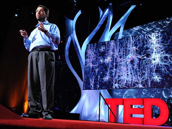
Humans have long held a fascination for the human brain. We chart it, we've described it, we've drawn it, we've mapped it. Now just like the physical maps of our world that have been highly influenced by technology -- think Google Maps, think GPS -- the same thing is happening for brain mapping through transformation.
İnsanlar çok uzun zamandır insan beynine hayran kalmıştır. Onun şemasını çıkardık, onu tanımladık, resimlerini çizdik ve haritasını çıkardık. Tıpkı dünyamızın teknolojiden büyük oranda etkilenen haritaları gibi -- Google Haritalar'ı, GPS'i düşünün -- aynı şey, dönüşümle beynin haritalanmasında da gerçekleşmektedir.
So let's take a look at the brain. Most people, when they first look at a fresh human brain, they say, "It doesn't look what you're typically looking at when someone shows you a brain." Typically, what you're looking at is a fixed brain. It's gray. And this outer layer, this is the vasculature, which is incredible, around a human brain. This is the blood vessels. 20 percent of the oxygen coming from your lungs, 20 percent of the blood pumped from your heart, is servicing this one organ. That's basically, if you hold two fists together, it's just slightly larger than the two fists.
Şimdi beyne bir göz atalım. Çoğu kimse, ilk kez taze bir insan beyni gördüğünde "Biri size bir beyin gösterdiğinde gördüğünüz şeye benzemiyor." der. Genellikle, gördüğünüz şey fikse edilmiş bir beyindir. Gri renktedir. Bu dış tabaka, insan beyninin etrafındaki, olağanüstü damar sistemidir. Bunlar kan damarlarıdır. Akciğerlerinizden gelen oksijenin yüzde 20'si, kalbinizden pompalanan kanın yüzde 20'si, yalnızca bu organa gitmektedir. İki yumruğunuzu yan yana koyarsanız, iki yumruğunuzdan biraz daha büyüktür.
Scientists, sort of at the end of the 20th century, learned that they could track blood flow to map non-invasively where activity was going on in the human brain. So for example, they can see in the back part of the brain, which is just turning around there. There's the cerebellum; that's keeping you upright right now. It's keeping me standing. It's involved in coordinated movement. On the side here, this is temporal cortex. This is the area where primary auditory processing -- so you're hearing my words, you're sending it up into higher language processing centers. Towards the front of the brain is the place in which all of the more complex thought, decision making -- it's the last to mature in late adulthood. This is where all your decision-making processes are going on. It's the place where you're deciding right now you probably aren't going to order the steak for dinner.
Bilimadamları, 20. yüzyılın sonlarında girişimsel olmayan yöntemlerle kan akışını takip ederek insan beyninin neresinde aktivite olduğunu haritalamanın mümkün olduğunu keşfettiler. Örneğin beynin arka tarafındaki aktiviteyi görebiliyorlar -- ekranda görmek üzere olduğunuz alan. İşte beyincik; şu anda sizi dik tutuyor. Benim ayakta durmamı sağlıyor. Hareket koordinasyonunda görev alır. Burada, yan tarafta, işte temporal korteks. Bu alanda birincil işitsel işlemler -- yani söylediklerimi duyuyorsunuz, yüksek dil işlem merkezine gönderiyorsunuz. Beynin ön tarafına doğru daha karmaşık düşüncelerin, karar verme işleminin gerçekleştiği -- erişkinlik döneminin sonlarında, en son olgunlaşandır. Burası tüm karar verme süreçlerinizin gerçekleştiği yerdir. Tam şu anda, büyük ihtimalle akşam yemeğinde biftek ısmarlamayacağınıza karar vermek için kullamakta olduğunuz bölge.
So if you take a deeper look at the brain, one of the things, if you look at it in cross-section, what you can see is that you can't really see a whole lot of structure there. But there's actually a lot of structure there. It's cells and it's wires all wired together. So about a hundred years ago, some scientists invented a stain that would stain cells. And that's shown here in the the very light blue. You can see areas where neuronal cell bodies are being stained. And what you can see is it's very non-uniform. You see a lot more structure there. So the outer part of that brain is the neocortex. It's one continuous processing unit, if you will. But you can also see things underneath there as well. And all of these blank areas are the areas in which the wires are running through. They're probably less cell dense. So there's about 86 billion neurons in our brain. And as you can see, they're very non-uniformly distributed. And how they're distributed really contributes to their underlying function. And of course, as I mentioned before, since we can now start to map brain function, we can start to tie these into the individual cells.
Beyne daha derinlemesine bakarsak, kesite bakarsak, fark edeceğiniz üzere burada pek fazla yapı görülemiyor. Ancak aslında çok fazla yapı mevcut. Tüm hücreleri ve tüm kabloları birbirine bağlanmış durumdadır. Yaklaşık yüz yıl kadar önce bilimadamları, hücreleri boyayan bir tür boya icat ettiler. Burada çok açık mavi renkte görülümektedir. Normal hücrelerin boyandığı alanları görebiliyoruz. Ve ne kadar tekdüze olmadığını görebiliyoruz. Burada çok daha fazla yapı görüyoruz. Beynin en dış bölümü neokortekstir. Tek parça, bütünleşik bir işlem birimi diyebiliriz. Fakat altında da bir şeyler görebiliyoruz. Tüm bu boş alanlar içinden kabloların geçtiği alanlardır. Büyük olasılıkla daha düşük hücre yoğunluğu mevcuttur. Beynimizde 86 milyar nöron vardır. Gördüğünüz üzere tekdüze olmayan bir şekilde dağılmışlardır. Dağılımlarının altta yatan fonksiyonlara bütük katkısı vardır. Tabii ki, daha önce de bahsettiğim üzere, artık beyin fonksiyonlarının haritasını çıkarmaya başlayabildiğimize göre bu fonksiyonları tek tek hücrelerle ilişkilendirebiliriz.
So let's take a deeper look. Let's look at neurons. So as I mentioned, there are 86 billion neurons. There are also these smaller cells as you'll see. These are support cells -- astrocytes glia. And the nerves themselves are the ones who are receiving input. They're storing it, they're processing it. Each neuron is connected via synapses to up to 10,000 other neurons in your brain. And each neuron itself is largely unique. The unique character of both individual neurons and neurons within a collection of the brain are driven by fundamental properties of their underlying biochemistry. These are proteins. They're proteins that are controlling things like ion channel movement. They're controlling who nervous system cells partner up with. And they're controlling basically everything that the nervous system has to do.
Şimdi biraz daha derinlemesine bakalım. Nöronlara bakalım. Daha önce de belirttiğim üzere, 86 milyar nöron var. Aynı zamanda, göreceğiniz üzere, daha ufak hücreler de var. Bunlar destek hücreleridir -- astrosit glia. Uyarıyı alanlar sinirlerin kendileridir. Uyarıyı depoluyorlar, işliyorlar. Her bir nöron, sinapslar aracılığıyla 10.000'e kadar nörona birden bağlı olabilmektedir. Her bir nöron büyük ölçüde özgündür. Hem beyindeki bir topluluğa dahil olarak, hem de tek başlarına ele alındıklarına, nöronların özgün karakterleri altta yatan biyokimyalarının temel özellikleri tarafından şekillenir. Bunlar proteinlerdir. İyon kanal hareketleri gibi şeyleri kontrol eden proteinlerdir. Sinir sistemi hücrelerinin ne ile ortaklaşa çalışacağını kontrol ederler. Temelde bu proteinler sinir sisteminin yapması gereken her şeyi kontrol ederler.
So if we zoom in to an even deeper level, all of those proteins are encoded by our genomes. We each have 23 pairs of chromosomes. We get one from mom, one from dad. And on these chromosomes are roughly 25,000 genes. They're encoded in the DNA. And the nature of a given cell driving its underlying biochemistry is dictated by which of these 25,000 genes are turned on and at what level they're turned on.
Daha derinlere doğru inersek bütün bu proteinler genomlarımız tarafından kodlanırlar. Her birimizin 23 çift kromozomu var. Bir çifti annemizden, diğerini babamızdan alırız. Bu kromozomlarda yaklaşık 25.000 gen var. DNA'da kodlanmışlardır. Herhangi bir hücrenin temel biyokimyasını belirleyen etkenler bu 25.000 genin hangilerinin ve hangi seviyede aktive olduğu ile belirlenir.
And so our project is seeking to look at this readout, understanding which of these 25,000 genes is turned on. So in order to undertake such a project, we obviously need brains. So we sent our lab technician out. We were seeking normal human brains. What we actually start with is a medical examiner's office. This a place where the dead are brought in. We are seeking normal human brains. There's a lot of criteria by which we're selecting these brains. We want to make sure that we have normal humans between the ages of 20 to 60, they died a somewhat natural death with no injury to the brain, no history of psychiatric disease, no drugs on board -- we do a toxicology workup. And we're very careful about the brains that we do take. We're also selecting for brains in which we can get the tissue, we can get consent to take the tissue within 24 hours of time of death. Because what we're trying to measure, the RNA -- which is the readout from our genes -- is very labile, and so we have to move very quickly.
Projemizde bu sonuçlara bakarak 25.000 genin hangilerinin aktive olduğunu anlamayı amaçlıyoruz. Böyle bir proje için doğal olarak beyinlere ihtiyacımız var. Biz de laboratuar teknisyenimizi yolladık. Normal insan beyni arıyoruz. Aslında adli tabibin çalışma odası ile başladık. Burası ölen insanların getirildiği yer. Normal insan beyni arıyoruz. Beyinleri seçmede kullandığımız bir çok ölçüt var. 20 ila 60 yaş arası doğal bir nedenden ölmüş beynine hiç zarar gelmemiş psikiyatrik hastalık geçmişi olmayan hiç narkotik ilaç kullanmamış -- -- toksikolojik tetkik yapıyoruz -- normal insanlar olduğundan emin olmak istiyoruz. Aldığımız beyinlerle çok dikkatli oluyoruz. Ayrıca, ölümü takiben 24 saat içinde dokuyu alabileceğimiz, dokuyu almak için izin alabileceğimiz beyinleri seçiyoruz. Çünkü ölçmeye çalıştığımız şey, yani RNA -- genlerimizin çıktısı -- son derece kararsızdır, dolayısıyla hızlı hareket etmemiz gerekiyor.
One side note on the collection of brains: because of the way that we collect, and because we require consent, we actually have a lot more male brains than female brains. Males are much more likely to die an accidental death in the prime of their life. And men are much more likely to have their significant other, spouse, give consent than the other way around.
Beyinlerin toplanmasına dair ufak bir not: toplama yönetimimiz ve rıza gerekliliği nedeniyle, elimizde kadın beyninden çok erkek beyni mevcut. Erkeklerin, hayatlarının en aktif zamanlarında bir kaza sonucu ölme ihtimalleri çok daha yüksektir. Ve erkeklerin, sevgili veya eşlerinin rızalarının alınması tam tersine göre daha olasıdır.
(Laughter)
(Kahkahalar)
So the first thing that we do at the site of collection is we collect what's called an MR. This is magnetic resonance imaging -- MRI. It's a standard template by which we're going to hang the rest of this data. So we collect this MR. And you can think of this as our satellite view for our map. The next thing we do is we collect what's called a diffusion tensor imaging. This maps the large cabling in the brain. And again, you can think of this as almost mapping our interstate highways, if you will. The brain is removed from the skull, and then it's sliced into one-centimeter slices. And those are frozen solid, and they're shipped to Seattle. And in Seattle, we take these -- this is a whole human hemisphere -- and we put them into what's basically a glorified meat slicer. There's a blade here that's going to cut across a section of the tissue and transfer it to a microscope slide. We're going to then apply one of those stains to it, and we scan it. And then what we get is our first mapping.
Beyni alacağımız yerde yaptığımız ilk iş MR çekmek oluyor. Manyetik rezonans görüntüleme -- MRG. Bu MR görüntüsü, geri kalan tüm verileri üzerine yerleştireceğimiz bir şablon görevi görüyor. MR çekiyoruz. Bunu haritamızın bir uydu görüntüsü olarak düşünebilirsiniz. Bir sonraki adımımız difüzyon tensör görüntülemesidir. Bu beyindeki büyük kabloların bir harıtasıdır. Bunu da şehirlerarası geniş yolların haritasını çıkarmak olarak düşünebilirsiniz. Beyin kafatasından çıkarılır ve bir santimetrelik dilimlere ayrılır. Bu dilimler kaskatı dondurulur ve Seattle'a gönderilir. Seatlle'da bu dilimleri alıp -- bu bütün bir insan beyninin yarısıdır (hemisfer) -- bir anlamda "kaliteli bir et dilimleme makinesi"ne koyuyoruz. Bu makinedeki bıçak, bir doku bölümünü boydan boya keser ve bir lama aktarır. Daha sonra o boyalardan birini uygulayıp tarama yapacağız. Böylece ilk haritamızı elde etmiş oluyoruz.
So this is where experts come in and they make basic anatomic assignments. You could consider this state boundaries, if you will, those pretty broad outlines. From this, we're able to then fragment that brain into further pieces, which then we can put on a smaller cryostat. And this is just showing this here -- this frozen tissue, and it's being cut. This is 20 microns thin, so this is about a baby hair's width. And remember, it's frozen. And so you can see here, old-fashioned technology of the paintbrush being applied. We take a microscope slide. Then we very carefully melt onto the slide. This will then go onto a robot that's going to apply one of those stains to it. And our anatomists are going to go in and take a deeper look at this.
Bu noktada uzmanlar devreye giriyor ve temel anatomik hatları belirliyorlar. Bu geniş hatları, eyalet sınırları olarak düşünebilirsiniz. Bu belirlemelere göre beyni, daha ufak kriyostatlara koymak üzere daha küçük parçalara ayırabiliyoruz. Ekranda da gördüğünüz üzere -- donmuş bir doku kesilmekte. Kesit kalınlığı 20 mikron, yani bir bebeğin saç teli kalınlığında. Unutmayın, doku donmuş durumda. Burada da gördüğünüz üzere eski bir teknoloji olan boya fırçası tekniği uygulanıyor. Bir lam alıyoruz. Çok dikkatli bir şekilde eriterek lamın üzerine koyuyoruz. Daha sonra dokunun üzerinde bulunduğu lama bir robot aracılığıyla boya uygulanacak. Sonra anatomi uzmanlarımız daha detaylı inceleme yapacak.
So again this is what they can see under the microscope. You can see collections and configurations of large and small cells in clusters and various places. And from there it's routine. They understand where to make these assignments. And they can make basically what's a reference atlas. This is a more detailed map.
Bu da mikroskop altında gördükleri görüntüdür. Kümeler içinde, çeşitli bölgelerde büyük ve küçük hücre toplulukları ve düzeni görüyoruz. Bu noktadan sonrası rutin oluyor. Nereleri belirlemeleri gerektiğini biliyorlar. Yani bir referans atlası oluşturabiliyorlar. Bu daha detaylı bir harita oluyor.
Our scientists then use this to go back to another piece of that tissue and do what's called laser scanning microdissection. So the technician takes the instructions. They scribe along a place there. And then the laser actually cuts. You can see that blue dot there cutting. And that tissue falls off. You can see on the microscope slide here, that's what's happening in real time. There's a container underneath that's collecting that tissue. We take that tissue, we purify the RNA out of it using some basic technology, and then we put a florescent tag on it. We take that tagged material and we put it on to something called a microarray.
Daha sonra bilimadamlarımız dokunun başka bir parçasına geçip lazer yardımlı mikrodiseksiyon denen tekniği uyguluyorlar. Teknisyen talimatları alır. Bu şekilde işaretleme yapar. Sonra da lazer, kesme işlemini gerçekleştirir. Orada mavi noktanın olduğu yerin kesildiğini görebilirsiniz. Kesilen doku aşağı düşer. Lamın üzerinde gördüğünüz gerçek zamanlıdır. Kesilen doku aşağıda duran bir toplama kabına düşer. O dokuyu alıp basit bir teknik kullanarak içindeki RNA'yı arıtır ve floresan işaretleme yaparız. İşaretlenmiş maddeyi alır ve mikrodizi denen bir şeye koyarız.
Now this may look like a bunch of dots to you, but each one of these individual dots is actually a unique piece of the human genome that we spotted down on glass. This has roughly 60,000 elements on it, so we repeatedly measure various genes of the 25,000 genes in the genome. And when we take a sample and we hybridize it to it, we get a unique fingerprint, if you will, quantitatively of what genes are turned on in that sample.
Şimdi, size yalnızca bir sürü nokta gibi gözüküyor olabilir. Fakat bu noktaların her biri aslında cam üzerinde saptadığımız özgün birer insan genomu parçasıdır. Burada yaklaşık 60.000 öğe var ve biz de, genomdaki 25.000 gen içinden çeşitli genleri tekrar tekrar ölçeriz. Bir numune alıp hibridize ettiğimizde numune içinde hangi genlerin aktive olduğunun sayısal olarak, özgün bir parmak izini elde ederiz.
Now we do this over and over again, this process for any given brain. We're taking over a thousand samples for each brain. This area shown here is an area called the hippocampus. It's involved in learning and memory. And it contributes to about 70 samples of those thousand samples. So each sample gets us about 50,000 data points with repeat measurements, a thousand samples.
Bu işlemi elimizdeki tüm beyinler için sürekli tekrarlıyoruz. Her bir beyinden binin üzerinde numune alıyoruz. Burada gösterilen bölgeye hipokampus deniyor. Bu bölge öğrenme ve hafıza işlemlerinde görev alır. Ve bu bölge, topladığımız bin numunenin yalnızca 70 kadarını oluşturur. Her bir numune bize 50.000 veri göstergesi sağlıyor ve tekrarlanan ölçümlerle, bin numune.
So roughly, we have 50 million data points for a given human brain. We've done right now two human brains-worth of data. We've put all of that together into one thing, and I'll show you what that synthesis looks like. It's basically a large data set of information that's all freely available to any scientist around the world. They don't even have to log in to come use this tool, mine this data, find interesting things out with this. So here's the modalities that we put together. You'll start to recognize these things from what we've collected before. Here's the MR. It provides the framework. There's an operator side on the right that allows you to turn, it allows you to zoom in, it allows you to highlight individual structures.
Yani bir insan beyni için elimizde yaklaşık 50 milyon veri göstergesi oluyor. Şu ana kadar iki insan beyni değerinde veri topladık. Tüm bu verileri tek bir şeyde topladık ve ortaya çıkan sentezin nasıl bir şey olduğunu size göstereyim. Dünyanın her yerindeki bilimadamlarına ücretsiz olarak açık geniş bir veri bankası. Bu aracı kullanmak, veri toplamak, ilginç şeyler keşfetmek için giriş bile yapmaları gerekmiyor. İşte bir araya getirdiğimiz araç. Önceden topladığımız şeyleri fark edeceksiniz. İşte MR görüntüsü. Ana iskeleti oluşturuyor. Sağ tarafta görüntüyü döndürmenize, yakınlaştırmanıza, her bir yapıyı tek tek seçmenize olanak sağlayan bir kontrol paneli var.
But most importantly, we're now mapping into this anatomic framework, which is a common framework for people to understand where genes are turned on. So the red levels are where a gene is turned on to a great degree. Green is the sort of cool areas where it's not turned on. And each gene gives us a fingerprint. And remember that we've assayed all the 25,000 genes in the genome and have all of that data available.
Fakat en önemlisi şimdi, insanların genlerin nerede aktive olduğunu anlayabilecekleri genel bir iskelet olan bu anatomik iskeleti haritalandırıyoruz. Kırmızı renkler bir genin yüksek seviyede aktive olduğu yerler. Yeşik renkler ise o genin aktive olmadığı soğuk bölgeler. Her bir gen bize bir parmak izi veriyor. Unutmayalım ki genomdaki 25.000 genin her birini çözümledik ve tüm veriler elimizde mevcut.
So what can scientists learn about this data? We're just starting to look at this data ourselves. There's some basic things that you would want to understand. Two great examples are drugs, Prozac and Wellbutrin. These are commonly prescribed antidepressants. Now remember, we're assaying genes. Genes send the instructions to make proteins. Proteins are targets for drugs. So drugs bind to proteins and either turn them off, etc. So if you want to understand the action of drugs, you want to understand how they're acting in the ways you want them to, and also in the ways you don't want them to. In the side effect profile, etc., you want to see where those genes are turned on. And for the first time, we can actually do that. We can do that in multiple individuals that we've assayed too.
Peki, bilimadamları bu veriler hakkında ne öğrenebilir? Biz de bu verilere yeni yeni bakmaya başladık. Anlamak isteyebileceğiniz iki ana şey var. İki önemli örnek ilaçlardır; Prozac ve Wellbutrin. Bunlar sıklıkla reçetelendirilen antidepresanlardır. Şimdi, genleri çözümlediğimizi unutmayalım. Genler, protein yapımı için gerekli talimatları gönderirler. Proteinler ilaçlar için birer hedeftir. Yani ilaçlar proteinlere bağlanır ve de-aktive ederler, vs. Bu durumda, ilaçların etkilerini anlamak istiyorsanız neden istediğiniz şekilde etki gösterdiklerini ve göstermediklerini anlamanız gerekir. Yan etki profili gibi şeylerde o genlerin nerede aktive olduğunu görmek istersiniz. İlk defa bunu gerçekten yapabiliyoruz. Bunu çözümlediğimiz diğer bireylerde de yapabiliyoruz.
So now we can look throughout the brain. We can see this unique fingerprint. And we get confirmation. We get confirmation that, indeed, the gene is turned on -- for something like Prozac, in serotonergic structures, things that are already known be affected -- but we also get to see the whole thing. We also get to see areas that no one has ever looked at before, and we see these genes turned on there. It's as interesting a side effect as it could be. One other thing you can do with such a thing is you can, because it's a pattern matching exercise, because there's unique fingerprint, we can actually scan through the entire genome and find other proteins that show a similar fingerprint. So if you're in drug discovery, for example, you can go through an entire listing of what the genome has on offer to find perhaps better drug targets and optimize.
Yani şimdi, beynin içine bütünüyle bakabiliyoruz. Bu özgün parmak izini görebiliyoruz. Ve doğrulayabiliyoruz. Gerçekten genin aktive olduğunu doğrulayabiliyoruz -- Prozac gibi bir ilaç için, serotonerjik yapılarda, etkilendiğini bildiğimiz şeyler için -- ama aynı zamanda herşeyi bütünüyle görebiliyoruz. Daha önce kimsenin bakmamış olduğu bölgelere bakabiliyor ve o bölgelerde bu genlerin aktive olduğunu görebiliyoruz. Bir yan etkinin olabildiği kadar ilginç. Bununla yapabileceğimiz bir başka şey ise, bu bir patern eşleştirme egzersizi olduğundan, özgün bir parmak izi olduğundan, tüm genomu tarayıp benzer parmak izi olan başka proteinler de bulabiliyoruz. Örneğin ilaç keşfi üzerine çalışıyorsanız, belki de tüm genom listesine bakıp daha iyi ilaç hedefleri bulabilir ve kullanımı optimize edebilirsiniz.
Most of you are probably familiar with genome-wide association studies in the form of people covering in the news saying, "Scientists have recently discovered the gene or genes which affect X." And so these kinds of studies are routinely published by scientists and they're great. They analyze large populations. They look at their entire genomes, and they try to find hot spots of activity that are linked causally to genes. But what you get out of such an exercise is simply a list of genes. It tells you the what, but it doesn't tell you the where. And so it's very important for those researchers that we've created this resource. Now they can come in and they can start to get clues about activity. They can start to look at common pathways -- other things that they simply haven't been able to do before.
Birçoğunuz, tüm genomu kapsayan ilişkilendirme çalışmaları sayesinde ortaya çıkan "Bilimadamları A'yı etkileyen gen veya genleri keşfetti." gibi haberleri görüyorsunuzdur. Bu tür çalışmalar bilimadamları tarafından düzenli olarak yayınlanıyor ve bu yayınlar çok güzel. Geniş popülasyonları analiz ediyorlar. Tüm genomlarına bakıyor ve nedensel olarak genlere bağlı yoğun aktivite bölgeleri bulmaya çalışıyorlar. Ancak bu tür bir çalışmadan elde edebileceğiniz şey en basitinden bir gen listesidir. Size ne olduğunu gösteriyor ama nerede olduğunu göstermiyor. Dolayısıyla bu kaynağı yaratmış olmamız o araştırmacılar için çok önemli. Şimdi bu kaynak ile aktiviteler hakkında bir fikir edinmeye başlayabilirler. Daha önce bakamadıkları yollarla genel yolaklara bakabilirler.
So I think this audience in particular can understand the importance of individuality. And I think every human, we all have different genetic backgrounds, we all have lived separate lives. But the fact is our genomes are greater than 99 percent similar. We're similar at the genetic level. And what we're finding is actually, even at the brain biochemical level, we are quite similar. And so this shows it's not 99 percent, but it's roughly 90 percent correspondence at a reasonable cutoff, so everything in the cloud is roughly correlated. And then we find some outliers, some things that lie beyond the cloud. And those genes are interesting, but they're very subtle. So I think it's an important message to take home today that even though we celebrate all of our differences, we are quite similar even at the brain level.
Sanırım özellikle buradaki dinleyiciler bireyselliğin önemini anlayabilirler. Bence her insan -- hepimizin genetik alt yapısı farklı, hepimizin ayrı hayatları var. Aslında genomlarımız yüzde 99'dan daha yüksek bir oranda benzerdir. Genetik seviyede benzeriz. Şimdi ise, aslında beyin biyokimyası seviyesinde bile çok benzer olduğumuzu anlıyoruz. Bu da yüzde 99 olmadığını, mantıklı bir ayırma noktası ile yaklaşık yüzde 90 olduğunu ve bulutun içindeki herşeyin yaklaşık olarak ilişkili olduğunu gösteriyor. Sonra, bulutun ötesinde bulunan bazı aykırı değerler buluyoruz. Bu genler ilginç ama hemen göze çarpmıyorlar. Bugün öğrendiğimiz önemli bir şey varsa o da her ne kadar tüm farklılıklarımızı vurguluyor olsak da aslında beyin seviyesinde bile çok benzer olduğumuzdur.
Now what do those differences look like? This is an example of a study that we did to follow up and see what exactly those differences were -- and they're quite subtle. These are things where genes are turned on in an individual cell type. These are two genes that we found as good examples. One is called RELN -- it's involved in early developmental cues. DISC1 is a gene that's deleted in schizophrenia. These aren't schizophrenic individuals, but they do show some population variation. And so what you're looking at here in donor one and donor four, which are the exceptions to the other two, that genes are being turned on in a very specific subset of cells. It's this dark purple precipitate within the cell that's telling us a gene is turned on there. Whether or not that's due to an individual's genetic background or their experiences, we don't know. Those kinds of studies require much larger populations.
Şimdi, bu farklılıklar neye benziyor? Bu, o farklılıkların tam olarak ne olduğunu görebilmek ve takip edebilmek için yaptığımız bir çalışmadan bir örnek -- ve o farklılıklar hiç göze batmayacak türde. Bunlar, belli bir hücre tipinde genlerin aktive olduğu yerler. Bulduğumuz bu iki genin iyi birer örnek olduğunu düşünüyoruz. Birine RELN adı veriliyor -- erken gelişim tetiklerinde görev alıyor. DISC1 şizofrenide silinen bir gendir. Bunlar şizofrenik bireyler değiller ama bazı popülasyon varyasonları sergiliyorlar. Burada donör bir ve donör dörtte gördüğünüz, diğer ikisine göre istisna olan şey, genlerin çok spesifik bir hücre altkümesinde aktive olduğudur. Burada bize genin aktive olduğunu gösteren şey, hücrenin içindeki koyu mor çökeltidir. Bunun, bireyin genetik alt yapısına veya deneyimlerine bağlı olup olmadığını bilmiyoruz. O tür çalışmalar çok daha geniş popülasyon gerektirir.
So I'm going to leave you with a final note about the complexity of the brain and how much more we have to go. I think these resources are incredibly valuable. They give researchers a handle on where to go. But we only looked at a handful of individuals at this point. We're certainly going to be looking at more. I'll just close by saying that the tools are there, and this is truly an unexplored, undiscovered continent. This is the new frontier, if you will. And so for those who are undaunted, but humbled by the complexity of the brain, the future awaits.
Size beynin karmaşıklığına ve daha ne kadar çok yolumuz olduğuna ilişkin bir not ile veda etmek istiyorum. Bence bu kaynaklar olağanüstü derece değerli. Araştırmacılara nereye yöneleceklerini gösteriyor. Fakat şu ana kadar incelediğimiz bireylerin sayısı çok az. Kesinlikle daha fazla bireyi inceliyor olacağız. Araçların elimizin altında olduğunu ve bu konunun gerçekten keşfedilmemiş, ortaya çıkarılmamış bir kıta olduğunu söylerek konuşmamı sonlandırmak istiyorum. Bu yeni sınırlarımızdır. Beynin karmaşıklığı karşısında mütevazı ama cesur olanlar; gelecek sizleri bekliyor.
Thanks.
Teşekkürler.
(Applause)
(Alkışlar)