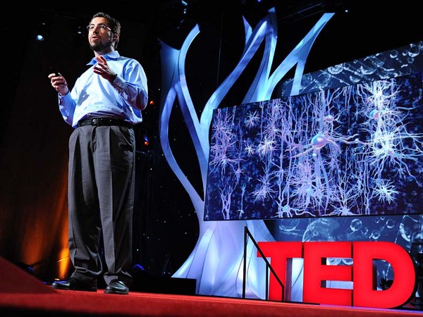
Humans have long held a fascination for the human brain. We chart it, we've described it, we've drawn it, we've mapped it. Now just like the physical maps of our world that have been highly influenced by technology -- think Google Maps, think GPS -- the same thing is happening for brain mapping through transformation.
Há tempos os homens têm uma fascinação pelo cérebro humano. Nós o traçamos, nós o descrevemos, nós o desenhamos, nós o mapeamos. Da mesma forma como os mapas físicos de nosso mundo que foram muito influenciados pela tecnologia -- pensem nos Google Maps, pensem no GPS -- a mesma coisa está acontecendo no mapeamento cerebral por meio de transformação.
So let's take a look at the brain. Most people, when they first look at a fresh human brain, they say, "It doesn't look what you're typically looking at when someone shows you a brain." Typically, what you're looking at is a fixed brain. It's gray. And this outer layer, this is the vasculature, which is incredible, around a human brain. This is the blood vessels. 20 percent of the oxygen coming from your lungs, 20 percent of the blood pumped from your heart, is servicing this one organ. That's basically, if you hold two fists together, it's just slightly larger than the two fists.
Então vamos dar uma olhada no cérebro. A maioria das pessoas, ao olhar pela primeira vez para um cérebro humano fresco, dizem: "Isso não parece com o que você vê quando alguém mostra um cérebro." Normalmente, o que vocês veem é um cérebro fixado. É cinza. E essa camada externa, é a vasculatura, que é incrível, ao redor do cérebro humano. Esses são os vasos sanguíneos. 20 por cento do oxigênio que vêm de seus pulmões, 20 por cento do sangue bombeado pelo seu coração, estão servindo a esse único órgão. Basicamente, se você apertar dois punhos juntos, ele será um pouco maior do que esses dois punhos.
Scientists, sort of at the end of the 20th century, learned that they could track blood flow to map non-invasively where activity was going on in the human brain. So for example, they can see in the back part of the brain, which is just turning around there. There's the cerebellum; that's keeping you upright right now. It's keeping me standing. It's involved in coordinated movement. On the side here, this is temporal cortex. This is the area where primary auditory processing -- so you're hearing my words, you're sending it up into higher language processing centers. Towards the front of the brain is the place in which all of the more complex thought, decision making -- it's the last to mature in late adulthood. This is where all your decision-making processes are going on. It's the place where you're deciding right now you probably aren't going to order the steak for dinner.
Os cientistas, perto do fim do século 20, aprenderam que podiam rastrear o fluxo sanguíneo para mapear de forma não invasiva onde a atividade estava ocorrendo no cérebro humano. Então por exemplo, eles podem ver na parte de trás do cérebro, que está virando aqui. Eis o cerebelo. Isso que está mantendo vocês sentados agora. Ele está me mantendo de pé. Está envolvido em movimentos coordenados. Aqui do lado, está o córtex temporal. Essa é a área onde ocorre o processamento auditivo primário -- então vocês escutam minhas palavras, e as enviam para centros de processamento de linguagem superiores. Em direção à frente do cérebro está o lugar de todos os pensamentos mais complexos, tomada de decisão -- é o último a amadurecer na fase adulta. É aqui onde todos os processos de tomada de decisão acontecem. É o lugar onde você decide agora mesmo se você vai pedir bife para o jantar.
So if you take a deeper look at the brain, one of the things, if you look at it in cross-section, what you can see is that you can't really see a whole lot of structure there. But there's actually a lot of structure there. It's cells and it's wires all wired together. So about a hundred years ago, some scientists invented a stain that would stain cells. And that's shown here in the the very light blue. You can see areas where neuronal cell bodies are being stained. And what you can see is it's very non-uniform. You see a lot more structure there. So the outer part of that brain is the neocortex. It's one continuous processing unit, if you will. But you can also see things underneath there as well. And all of these blank areas are the areas in which the wires are running through. They're probably less cell dense. So there's about 86 billion neurons in our brain. And as you can see, they're very non-uniformly distributed. And how they're distributed really contributes to their underlying function. And of course, as I mentioned before, since we can now start to map brain function, we can start to tie these into the individual cells.
Então se você der uma olhada mais profunda no cérebro uma das coisas, se você observá-lo nessa seção, o que você pode ver é que não pode realmente ver um monte de estruturas aqui. Mas há realmente um monte de estruturas aqui. Suas células e suas conexões estão todas ligadas. Então, cerca de 100 anos atrás, alguns cientistas inventaram uma marcação que podia identificar células. E isso é mostrado aqui nesse azul bem claro. Você pode ver áreas onde corpos celulares normais estão marcados. E você pode ver que não é muito uniforme. Você vê um monte de estruturas ali. Então a parte externa desse cérebro é o neocórtex. É uma unidade de processamento contínuo, se preferir. Mas vocês também podem ver coisas embaixo dele. Todas essas áreas em branco são as áreas onde as conexões estão passando. Elas provavelmente são menos densas em células. Então há cerca de 86 billhões de neurônios em nosso cérebro. E como pode ver aqui, eles não estão distribuídos uniformemente. E como eles se distribuem contribui muito para sua função subjacente. E é claro, como mencionei antes, como podemos agora começar a mapear a função cerebral, podemos começar a amarrá-las em células individuais.
So let's take a deeper look. Let's look at neurons. So as I mentioned, there are 86 billion neurons. There are also these smaller cells as you'll see. These are support cells -- astrocytes glia. And the nerves themselves are the ones who are receiving input. They're storing it, they're processing it. Each neuron is connected via synapses to up to 10,000 other neurons in your brain. And each neuron itself is largely unique. The unique character of both individual neurons and neurons within a collection of the brain are driven by fundamental properties of their underlying biochemistry. These are proteins. They're proteins that are controlling things like ion channel movement. They're controlling who nervous system cells partner up with. And they're controlling basically everything that the nervous system has to do.
Então vamos dar uma olhada mais profunda. Vamos olhar os neurônios. Como mencionei, há 86 bilhões de neurônios. Há também essas células menores como podem ver. Elas são células de suporte -- astrócitos da glia. E os próprios nervos são aqueles que recebem aferências. Eles estão armazenando, eles estão processando. Cada neurônio está conectado via sinapses com até 10 mil outros neurônios em seu cérebro. E cada neurônio sozinho é muito singular. O caráter singular tanto de neurônios individuais como de neurônios dentro de um aglomerado cerebral é determinada por propriedades fundamentais de sua bioquímica subjacente. Essas são proteínas. São proteínas que controlam coisas como movimento de canais iônicos. Elas controlam com quem as células do sistema nervoso se conectam. E elas controlam basicamente tudo o que o sistema nervoso precisa fazer.
So if we zoom in to an even deeper level, all of those proteins are encoded by our genomes. We each have 23 pairs of chromosomes. We get one from mom, one from dad. And on these chromosomes are roughly 25,000 genes. They're encoded in the DNA. And the nature of a given cell driving its underlying biochemistry is dictated by which of these 25,000 genes are turned on and at what level they're turned on.
Então se focarmos para um nível ainda mais profundo, todas essas proteínas são codificadas por nossos genomas. Cada um de nós tem 23 pares de cromossomos. Nós recebemos um da mãe e um do pai. E nesses cromossomos estão aproximadamente 25 mil genes. Eles estão codificados no DNA. E a natureza de uma determinada célula que dirige sua bioquímica subjacente é ditada por quais desses 25 mil genes são ativados e em qual nível estão ativados.
And so our project is seeking to look at this readout, understanding which of these 25,000 genes is turned on. So in order to undertake such a project, we obviously need brains. So we sent our lab technician out. We were seeking normal human brains. What we actually start with is a medical examiner's office. This a place where the dead are brought in. We are seeking normal human brains. There's a lot of criteria by which we're selecting these brains. We want to make sure that we have normal humans between the ages of 20 to 60, they died a somewhat natural death with no injury to the brain, no history of psychiatric disease, no drugs on board -- we do a toxicology workup. And we're very careful about the brains that we do take. We're also selecting for brains in which we can get the tissue, we can get consent to take the tissue within 24 hours of time of death. Because what we're trying to measure, the RNA -- which is the readout from our genes -- is very labile, and so we have to move very quickly.
Então nosso projeto é tentar verificar esta leitura, e entender quais desses 25 mil genes estão ativados. Para executar um projeto assim, nós precisamos de cérebros, obviamente. Então enviamos nosso técnico de laboratório para fora. Nós estávamos em busca de cérebros humanos normais. Nós começamos na verdade em um instituto médico legal. Esse é um lugar para onde os mortos são trazidos. Nós estamos procurando cérebros humanos normais. Há vários critérios pelos quais selecionamos esses cérebros. Nós queremos ter certeza de que temos humanos normais entre as idades de 20 a 60 anos, que morreram devido a morte natural sem danos para o cérebro, sem histórico de doença psiquiátrica, sem drogas envolvidas -- nós fazemos um exame toxicológico. E somos muito cuidadosos com os cérebros que levamos. Nós também selecionamos cérebros em que podemos retirar o tecido, podemos conseguir o consentimento para retirar o tecido até 24 horas depois da hora da morte. Pois o que estamos tentando medir, o RNA -- que é a leitura de nossos genes -- é muito lábil, e por isso temos de agir com rapidez.
One side note on the collection of brains: because of the way that we collect, and because we require consent, we actually have a lot more male brains than female brains. Males are much more likely to die an accidental death in the prime of their life. And men are much more likely to have their significant other, spouse, give consent than the other way around.
Uma nota sobre a coleção de cérebros: devido à maneira como os coletamos, e por que pedimos consentimento, nós temos muito mais cérebros de homens do que de mulheres. Os homens tendem muito mais a morrer em acidentes no auge da vida. E os homens tendem muito mais a ter uma companheira, sua esposa, que dá o consentimento, do que o inverso.
(Laughter)
(Risos)
So the first thing that we do at the site of collection is we collect what's called an MR. This is magnetic resonance imaging -- MRI. It's a standard template by which we're going to hang the rest of this data. So we collect this MR. And you can think of this as our satellite view for our map. The next thing we do is we collect what's called a diffusion tensor imaging. This maps the large cabling in the brain. And again, you can think of this as almost mapping our interstate highways, if you will. The brain is removed from the skull, and then it's sliced into one-centimeter slices. And those are frozen solid, and they're shipped to Seattle. And in Seattle, we take these -- this is a whole human hemisphere -- and we put them into what's basically a glorified meat slicer. There's a blade here that's going to cut across a section of the tissue and transfer it to a microscope slide. We're going to then apply one of those stains to it, and we scan it. And then what we get is our first mapping.
Então a primeira coisa que fazemos no local da coleta é coletarmos o que chamamos de MR. Isso é imageamento por ressonância magnética -- MRI. É uma amostra padrão pela qual vamos basear o resto dos dados. Então coletamos esse MR. E você pode pensar nisso como nossa visão de satélite de nosso mapa. A próxima coisa que fazemos é coletar o que é chamado de imageamento por tensor de difusão. Isso mapeia os cabos principais do cérebro. E de novo, você pode pensar nisso como o mapeamento de nossas rodovias federais, se preferir. O cérebro é removido do crânio, e é então cortado em fatias de 1 centímetro. E elas são congeladas, e são enviadas para Seattle. E em Seattle, nós fazemos isso -- isso é um hemisfério humano inteiro -- e o colocamos num tipo de fatiador de carne especial. Há uma lâmina que corta uma seção do tecido e a transfere para uma lâmina de microscópio. Nós vamos aplicar uma das marcações nela, e a escaneamos. E depois o que temos é nosso primeiro mapeamento.
So this is where experts come in and they make basic anatomic assignments. You could consider this state boundaries, if you will, those pretty broad outlines. From this, we're able to then fragment that brain into further pieces, which then we can put on a smaller cryostat. And this is just showing this here -- this frozen tissue, and it's being cut. This is 20 microns thin, so this is about a baby hair's width. And remember, it's frozen. And so you can see here, old-fashioned technology of the paintbrush being applied. We take a microscope slide. Then we very carefully melt onto the slide. This will then go onto a robot that's going to apply one of those stains to it. And our anatomists are going to go in and take a deeper look at this.
Então aqui é onde os experts entram e fazem medidas anatômicas básicas. Você pode considerar como fronteiras entre os estados, se preferir, essas lindas linhas fronteiriças. A partir disso, somos capazes de fragmentar esse cérebro em peças menores, que colocamos em um criostato menor. E isso está mostrado aqui -- o tecido congelado, e sendo cortado. Isso tem 20 mícrons de espessura, mais ou menos um fio de cabelo de um bebê. E lembre-se, está congelado. E como podem ver aqui, a tradicional tecnologia do pincel sendo aplicada. Nós usamos uma lâmina de microscópio. Então nós a esquentamos com muito cuidado sobre a lâmina. Isso vai depois para um robô que aplica uma dessas marcações nela. E nossos anatomistas vão dar uma olhada mais profunda nisso.
So again this is what they can see under the microscope. You can see collections and configurations of large and small cells in clusters and various places. And from there it's routine. They understand where to make these assignments. And they can make basically what's a reference atlas. This is a more detailed map.
Então novamente isso é o que eles veem no microscópio. Vocês podem ver coleções e configurações de células maiores e menores em grupos e lugares variados. E a partir daí é rotina. Eles sabem onde fazer essas medidas. E eles podem fazer basicamente um atlas de referência. Esse é um mapa mais detalhado.
Our scientists then use this to go back to another piece of that tissue and do what's called laser scanning microdissection. So the technician takes the instructions. They scribe along a place there. And then the laser actually cuts. You can see that blue dot there cutting. And that tissue falls off. You can see on the microscope slide here, that's what's happening in real time. There's a container underneath that's collecting that tissue. We take that tissue, we purify the RNA out of it using some basic technology, and then we put a florescent tag on it. We take that tagged material and we put it on to something called a microarray.
Nossos cientistas usam isso para voltar para outra peça desse tecido e fazer a chamada microdissecção por escaneamento a laser. Então os técnicos tomam as instruções. Eles delineiam um lugar ali. E depois o laser o corta. Vocês podem ver esse ponto azul cortando. E esse tecido cai. Vocês podem vê-lo na lâmina de microscópio aqui, isso é o que acontece em tempo real. Há um recipiente abaixo que está coletando esse tecido. Nós pegamos esse tecido, purificamos o RNA dele usando alguma tecnologia básica, e depois colocamos uma marca fluorescente nele. Nós pegamos esse material marcado e o colocamos em algo chamado de 'microarray'.
Now this may look like a bunch of dots to you, but each one of these individual dots is actually a unique piece of the human genome that we spotted down on glass. This has roughly 60,000 elements on it, so we repeatedly measure various genes of the 25,000 genes in the genome. And when we take a sample and we hybridize it to it, we get a unique fingerprint, if you will, quantitatively of what genes are turned on in that sample.
Agora isso pode parecer como um monte de pontos para vocês, mas cada um desses pontos individuais representa um pedaço singular do genoma humano que detectamos na lâmina. Esse tem cerca de 60 mil elementos, então nós medimos repetidamente vários genes dos 25 mil genes do genoma. E quando tomamos uma amostra e a hibridizamos, conseguimos uma digital singular, se preferir, quantitativa de quais genes estão ativados naquela amostra.
Now we do this over and over again, this process for any given brain. We're taking over a thousand samples for each brain. This area shown here is an area called the hippocampus. It's involved in learning and memory. And it contributes to about 70 samples of those thousand samples. So each sample gets us about 50,000 data points with repeat measurements, a thousand samples.
Agora fazemos isso de novo e de novo, esse processo para todos os cérebros. Estamos coletando mil amostras para cada cérebro. Essa área mostrada aqui é chamada de hipocampo. Ela está envolvida em aprendizagem e memória. E ela contribui com cerca de 70 amostras das mil amostras. Então cada amostra nos oferece cerca de 50 mil pontos de dados com medidas repetidas, mil amostras.
So roughly, we have 50 million data points for a given human brain. We've done right now two human brains-worth of data. We've put all of that together into one thing, and I'll show you what that synthesis looks like. It's basically a large data set of information that's all freely available to any scientist around the world. They don't even have to log in to come use this tool, mine this data, find interesting things out with this. So here's the modalities that we put together. You'll start to recognize these things from what we've collected before. Here's the MR. It provides the framework. There's an operator side on the right that allows you to turn, it allows you to zoom in, it allows you to highlight individual structures.
Então temos aproximadamente 50 milhões de pontos de dados para cada cérebro humano. Nós conseguimos agora dados completos de dois cérebros humanos. Nós juntamos tudo em uma coisa só, e vou mostrar a vocês como se parece a síntese. É basicamente uma grande série de informações que está disponível gratuitamente para qualquer cientista no planeta. Não é preciso fazer cadastro para usar essa ferramenta, analisar os dados, encontrar coisas interessantes com isso. Então eis as modalidades que colocamos juntas. Vocês vão reconhecer essas coisas das que já coletamos antes. Aqui está o MR. Ele fornece a estrutura. Há um operador lateral à direita que permite que você vire, faça um zoom, permite que você destaque estruturas individuais.
But most importantly, we're now mapping into this anatomic framework, which is a common framework for people to understand where genes are turned on. So the red levels are where a gene is turned on to a great degree. Green is the sort of cool areas where it's not turned on. And each gene gives us a fingerprint. And remember that we've assayed all the 25,000 genes in the genome and have all of that data available.
Mas mais importante, estamos agora mapeando essa estrutura anatômica, que é uma estrutura comum para as pessoas entenderem onde os genes estão ativados. Então os níveis em vermelho são onde um gene está ativado em um nível alto. O verde são as áreas frias onde não está ativado. E cada gene nos dá uma digital. E lembre-se que analisamos todos os 25 mil genes do genoma e temos todos esses dados disponíveis.
So what can scientists learn about this data? We're just starting to look at this data ourselves. There's some basic things that you would want to understand. Two great examples are drugs, Prozac and Wellbutrin. These are commonly prescribed antidepressants. Now remember, we're assaying genes. Genes send the instructions to make proteins. Proteins are targets for drugs. So drugs bind to proteins and either turn them off, etc. So if you want to understand the action of drugs, you want to understand how they're acting in the ways you want them to, and also in the ways you don't want them to. In the side effect profile, etc., you want to see where those genes are turned on. And for the first time, we can actually do that. We can do that in multiple individuals that we've assayed too.
Então o que os cientistas podem aprender com esses dados? Nós mesmos estamos só começando a olhar para esses dados. Há algumas coisas básicas que vocês gostariam de saber. Dois grandes exemplos são as drogas, Prozac e Wellbutrin. Eles são antidepressivos normalmente prescritos. Agora lembrem-se, estamos analisando genes. Os genes enviam as instruções para fazer proteínas. As proteínas são alvos para as drogas. Então as drogas se ligam a proteínas e as ativam, etc. Então se você quer entender a ação das drogas, é preciso entender como elas atuam nas maneiras que gostaríamos, e também nas maneiras que não gostaríamos. Seus efeitos colaterais, etc. Você quer ver onde esses genes são ativados. E pela primeira vez, nós podemos fazer isso. Podemos fazer isso em vários indivíduos que analisamos também.
So now we can look throughout the brain. We can see this unique fingerprint. And we get confirmation. We get confirmation that, indeed, the gene is turned on -- for something like Prozac, in serotonergic structures, things that are already known be affected -- but we also get to see the whole thing. We also get to see areas that no one has ever looked at before, and we see these genes turned on there. It's as interesting a side effect as it could be. One other thing you can do with such a thing is you can, because it's a pattern matching exercise, because there's unique fingerprint, we can actually scan through the entire genome and find other proteins that show a similar fingerprint. So if you're in drug discovery, for example, you can go through an entire listing of what the genome has on offer to find perhaps better drug targets and optimize.
Então agora podemos ver através do cérebro. Podemos ver essa digital única. E obter a confirmação. Obtemos a confirmação de que, de fato, o gene está ativado -- por algo como o Prozac, em estruturas serotonérgicas, coisas que já se sabia que são afetadas -- mas também conseguimos ver o quadro completo. Podemos ver áreas que ninguém havia olhado antes, e vemos esses genes ativados aqui. É um efeito colateral muito interessante. Uma outra coisa que você pode fazer com isso é que você pode, pois é um exercício de comparação de padrões, pois há uma digital singular, podemos escanear o genoma completo e encontrar outras proteínas que mostram uma digital similar. Então se você pesquisa a descoberta de novas drogas, por exemplo, você pode fazer uma lista completa do que o genoma tem a oferecer para encontrar talvez melhores drogas e otimizá-las.
Most of you are probably familiar with genome-wide association studies in the form of people covering in the news saying, "Scientists have recently discovered the gene or genes which affect X." And so these kinds of studies are routinely published by scientists and they're great. They analyze large populations. They look at their entire genomes, and they try to find hot spots of activity that are linked causally to genes. But what you get out of such an exercise is simply a list of genes. It tells you the what, but it doesn't tell you the where. And so it's very important for those researchers that we've created this resource. Now they can come in and they can start to get clues about activity. They can start to look at common pathways -- other things that they simply haven't been able to do before.
A maioria de vocês provavelmente conhece estudos de associação de genomas na forma como as pessoas acompanham as notícias dizendo: "Cientistas descobriram recentemente o gene ou genes que afetam X." E assim esse tipo de estudos são rotineiramente publicados pelos cientistas e são ótimos. Eles analisam grandes populações. Eles verificam genomas inteiros, e tentam encontrar pontos quentes de atividade que estão ligados causalmente com genes. Mas o que você obtém com um estudo assim é apenas uma lista de genes. Ele diz o que, mas não diz onde. Assim é muito importante para esses pesquisadores que tenhamos criado esse recurso. Agora eles podem vir e podem começar a ter pistas sobre atividade. Eles podem começar a olhar as vias comuns -- outros caminhos que simplesmente não podiam ver antes.
So I think this audience in particular can understand the importance of individuality. And I think every human, we all have different genetic backgrounds, we all have lived separate lives. But the fact is our genomes are greater than 99 percent similar. We're similar at the genetic level. And what we're finding is actually, even at the brain biochemical level, we are quite similar. And so this shows it's not 99 percent, but it's roughly 90 percent correspondence at a reasonable cutoff, so everything in the cloud is roughly correlated. And then we find some outliers, some things that lie beyond the cloud. And those genes are interesting, but they're very subtle. So I think it's an important message to take home today that even though we celebrate all of our differences, we are quite similar even at the brain level.
Então eu acho que essa audiência em particular pode entender a importância da individualidade. E eu acho que cada humano, todos nós temos contextos genéticos diferentes, todos nós vivemos vidas separadas. Mas o fato é que nossos genomas são mais do que 99 por cento parecidos. Nós somos parecidos no nível genético. E o que estamos descobrindo é que, mesmo no nível da bioquímica cerebral, nós somos muito parecidos. E isso mostra que não é 99 por cento, mas é uma correspondência de 90 por cento em um limite razoável, então tudo na nuvem está correlacionado aproximadamente. E depois encontramos alguns pontos fora da curva, algumas coisas que estão fora da nuvem. E esses genes são interessantes, mas eles são muito sutis. Então acho que é uma mensagem importante para levar para casa hoje que mesmo que celebremos todas as nossas diferenças, nós somos muito parecidos mesmo no nível cerebral.
Now what do those differences look like? This is an example of a study that we did to follow up and see what exactly those differences were -- and they're quite subtle. These are things where genes are turned on in an individual cell type. These are two genes that we found as good examples. One is called RELN -- it's involved in early developmental cues. DISC1 is a gene that's deleted in schizophrenia. These aren't schizophrenic individuals, but they do show some population variation. And so what you're looking at here in donor one and donor four, which are the exceptions to the other two, that genes are being turned on in a very specific subset of cells. It's this dark purple precipitate within the cell that's telling us a gene is turned on there. Whether or not that's due to an individual's genetic background or their experiences, we don't know. Those kinds of studies require much larger populations.
Agora como essas diferenças se parecem? Isso é um exemplo de um estudo que fizemos para rastrear e ver como eram essas diferenças exatamente -- e elas são muito sutis. Essas são coisas onde os genes são ativados em um tipo de célula específico. Esses são dois genes que encontramos como bons exemplos. Um é chamado de RELN -- está envolvido com pistas do desenvolvimento inicial. O DISC1 é um gene que está deletado na esquizofrenia. Esses não são indivíduos esquizofrênicos, mas eles mostram alguma variação na população. E o que vocês estão vendo aqui no doador um e doador quatro, que são as exceções dos outros dois, é que os genes estão sendo ativados em um grupo muito específico de células. É esse precipitado roxo escuro dentro da célula que está nos dizendo que um gene está ativado ali. Se isso é ou não é devido ao contexto genético do indivíduo ou suas experiências, nós não sabemos. Esses tipos de estudos requerem populações bem maiores.
So I'm going to leave you with a final note about the complexity of the brain and how much more we have to go. I think these resources are incredibly valuable. They give researchers a handle on where to go. But we only looked at a handful of individuals at this point. We're certainly going to be looking at more. I'll just close by saying that the tools are there, and this is truly an unexplored, undiscovered continent. This is the new frontier, if you will. And so for those who are undaunted, but humbled by the complexity of the brain, the future awaits.
Então vou deixar a vocês uma nota final sobre a complexidade do cérebro e quanto ainda precisamos ir. Eu acho que esses recursos são incrivelmente valiosos. Eles oferecem aos pesquisadores uma bússola para onde ir. Mas nós só olhamos para alguns indivíduos até esse ponto. Certamente vamos olhar em mais. Eu vou apenas encerrar dizendo que as ferramentas estão aí, e isso é realmente um continente inexplorado e desconhecido. Essa é a nova fronteira, se preferir. E para aqueles que são destemidos, mas humildes diante da complexidade do cérebro, o futuro aguarda.
Thanks.
Obrigado.
(Applause)
(Aplausos)