Humans have long held a fascination for the human brain. We chart it, we've described it, we've drawn it, we've mapped it. Now just like the physical maps of our world that have been highly influenced by technology -- think Google Maps, think GPS -- the same thing is happening for brain mapping through transformation.
인간은 아주 오랫동안 인간의 뇌의 신비에 매료되어 왔습니다. 우리는 뇌를 표로 그리고, 설명을 덧붙이고, 그림으로 표현하였으며, 지도로 만들었습니다. 이제 오늘날의 기술에 영향을 받아 변화하는 실제 지도들을 생각해보시죠 구글 지도를 떠올려 보시고, GPS 를 생각해 보세요 이런 기술의 변화가 두뇌 지도 생성에도 응용되고 있습니다.
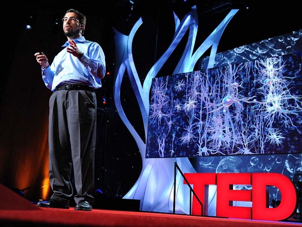
So let's take a look at the brain. Most people, when they first look at a fresh human brain, they say, "It doesn't look what you're typically looking at when someone shows you a brain." Typically, what you're looking at is a fixed brain. It's gray. And this outer layer, this is the vasculature, which is incredible, around a human brain. This is the blood vessels. 20 percent of the oxygen coming from your lungs, 20 percent of the blood pumped from your heart, is servicing this one organ. That's basically, if you hold two fists together, it's just slightly larger than the two fists.
자 이제 뇌를 관찰해 봅시다. 대부분의 사람들은 인간의 뇌를 실제로 보고나서 이렇게 말합니다, "이전에 보았던 두뇌와는 뭔가 달라 보여요." 보통, 여러분이 보아 왔던 두뇌는 멈춰 있는 두뇌입니다. 회색이죠. 그리고 이 바깥막은, 두뇌를 둘러싸고 있는 것은 맥관구조로서 대단한 존재입니다. 이것은 혈관들 입니다. 여러분의 폐에서 나온 20%의 산소와 여러분의 심장에서 만들어낸 20%의 혈액이 이 장기 한개를 지원하고 있습니다. 쉽게말해, 두 주먹을 쥐어보시면 뇌는 그 두 주먹보다 약간 더 큰 크기입니다.
Scientists, sort of at the end of the 20th century, learned that they could track blood flow to map non-invasively where activity was going on in the human brain. So for example, they can see in the back part of the brain, which is just turning around there. There's the cerebellum; that's keeping you upright right now. It's keeping me standing. It's involved in coordinated movement. On the side here, this is temporal cortex. This is the area where primary auditory processing -- so you're hearing my words, you're sending it up into higher language processing centers. Towards the front of the brain is the place in which all of the more complex thought, decision making -- it's the last to mature in late adulthood. This is where all your decision-making processes are going on. It's the place where you're deciding right now you probably aren't going to order the steak for dinner.
20세기 말경 과학자들은 혈액의 흐름을 비침범적으로 추적하여 여러 활동들이 이뤄지고 있는 두뇌 내부의 지도를 그릴 수 있다는 것을 발견 하였습니다. 예를 들어, 그들은 두뇌의 뒷 부분을 볼 수 있었으며 바로 이 부분이죠. 여러분이 똑바로 서 있을수 있게 도와주는 이것은 소뇌이며 조화로운 동작을 행할 수 있게 합니다. 이 옆부분은 측두엽 피질 입니다. 여기서 일차 청각 처리가 이뤄지게 되며 여러분들이 제 말을 듣고 난 뒤, 그것을 더 높은 수준의 중앙 언어처리 장치로 보내게 합니다. 두뇌의 앞 방향에서는 더 복잡한 생각과 결정을 내리는 부분이 위치하며 성인기 말에 마지막으로 성숙해지는 부분 입니다. 모든 의사 결정을 여기서 내립니다. 현재 여러분이 결정을 내리고 있는 부분이며 오늘 저녁엔 스테이크를 먹지 말아야지 라는 결정도 내립니다.
So if you take a deeper look at the brain, one of the things, if you look at it in cross-section, what you can see is that you can't really see a whole lot of structure there. But there's actually a lot of structure there. It's cells and it's wires all wired together. So about a hundred years ago, some scientists invented a stain that would stain cells. And that's shown here in the the very light blue. You can see areas where neuronal cell bodies are being stained. And what you can see is it's very non-uniform. You see a lot more structure there. So the outer part of that brain is the neocortex. It's one continuous processing unit, if you will. But you can also see things underneath there as well. And all of these blank areas are the areas in which the wires are running through. They're probably less cell dense. So there's about 86 billion neurons in our brain. And as you can see, they're very non-uniformly distributed. And how they're distributed really contributes to their underlying function. And of course, as I mentioned before, since we can now start to map brain function, we can start to tie these into the individual cells.
자 이제 더 깊게 두뇌를 들여다 보면 아시겠지만, 두뇌의 단면을 통하여 제대로 된 두뇌의 구조를 알아보기가 쉽지 않다는 것 입니다. 사실, 이곳에는 많은 조직들이 존재하고 있으며 세포들과 선들이 다같이 연결되어 있습니다. 그래서 약 100년 전, 몇몇 과학자들은 세포 염색제를 개발했습니다. 보시는 밝은 파란색이 그것입니다. 정상적인 세포기관들이 염색되는 부분들을 지금 보시고 계십니다. 매우 일관성이 없어 보이죠. 훨씬 더 많은 조직이 보입니다. 지금 보시는 두뇌의 바깥부분은 신피질 이라고 합니다. 지속적인 작업을 수행하는 단일체라고 생각하시면 됩니다. 하지만 그 아래에 있는 것들도 같이 볼 수 있습니다. 그리고 이 비어있는 영역들 역시 많은 것들이 연결되어 있는 영역들 입니다. 세포 밀도가 좀 더 낮겠죠. 인간의 뇌에는 약 860억개의 뉴런이 있습니다. 보시다시피, 매우 비균일적으로 분포되어 있으며 어떻게 분포되어 있는지가 그들의 기본적인 기능에 중요한 기여를 합니다. 그리고 물론, 제가 말했다시피, 뇌 기능을 지도로 그려내는 것이 가능하기에, 각각의 세포에 연관지어 볼 수 있습니다.
So let's take a deeper look. Let's look at neurons. So as I mentioned, there are 86 billion neurons. There are also these smaller cells as you'll see. These are support cells -- astrocytes glia. And the nerves themselves are the ones who are receiving input. They're storing it, they're processing it. Each neuron is connected via synapses to up to 10,000 other neurons in your brain. And each neuron itself is largely unique. The unique character of both individual neurons and neurons within a collection of the brain are driven by fundamental properties of their underlying biochemistry. These are proteins. They're proteins that are controlling things like ion channel movement. They're controlling who nervous system cells partner up with. And they're controlling basically everything that the nervous system has to do.
자 그럼 한단계 더 들어가 볼까요. 뉴런을 살펴봅시다. 말씀드렸다시피, 860억개의 뉴런이 존재하고 있으며 그보다 더 작은 세포들도 여러분은 보시게 될 것입니다. 아교세포 라고 불리는 지원세포들 입니다. 그리고 신경들 자체가 입력을 받고 있는 중이며 보관하고, 처리하는 작업도 합니다. 각 뉴런은 연접을 통해 두뇌의 최대 1만개 까지의 다른 뉴런들과 연결되어 있습니다. 그리고 각각의 뉴런은 매우 특이합니다. 각각의 뉴런들과 두뇌 안에 위치한 뉴런집합의 특성은 생화학 고유의 성질에 따라 행동하게 됩니다. 이것들은 단백질 입니다. 이 단백질들은 이온 채널의 움직임을 통제하며 신경시스템 세포와 누가 교신을 이루는가를 통제함과 더불어 다른 모든 신경 시스템이 하는일을 관장합니다.
So if we zoom in to an even deeper level, all of those proteins are encoded by our genomes. We each have 23 pairs of chromosomes. We get one from mom, one from dad. And on these chromosomes are roughly 25,000 genes. They're encoded in the DNA. And the nature of a given cell driving its underlying biochemistry is dictated by which of these 25,000 genes are turned on and at what level they're turned on.
자 여기서 한단계 더 깊은 레벨로 확대해서 보면, 이 모든 단백질이 각자의 게놈에 의하여 암호화 됩니다. 여러분은 각각 23쌍의 염색체를 가지고 있습니다. 우리는 한개는 어머니로부터, 한개는 아버지로부터 받습니다. 그리고 이 염색체에는 2만 5천개의 유전자가 존재하고 있습니다. DNA 속에 인식되어 있습니다. 그리고 기초 생화학 작용을 이끄는 세포의 본성은 2만 5천개의 유전자중 어떤 유전자가 반응하는가에 따라 그리고 어떤 레벨에서 반응하는지에 따라 정해지게 됩니다.
And so our project is seeking to look at this readout, understanding which of these 25,000 genes is turned on. So in order to undertake such a project, we obviously need brains. So we sent our lab technician out. We were seeking normal human brains. What we actually start with is a medical examiner's office. This a place where the dead are brought in. We are seeking normal human brains. There's a lot of criteria by which we're selecting these brains. We want to make sure that we have normal humans between the ages of 20 to 60, they died a somewhat natural death with no injury to the brain, no history of psychiatric disease, no drugs on board -- we do a toxicology workup. And we're very careful about the brains that we do take. We're also selecting for brains in which we can get the tissue, we can get consent to take the tissue within 24 hours of time of death. Because what we're trying to measure, the RNA -- which is the readout from our genes -- is very labile, and so we have to move very quickly.
그래서 우리의 프로젝트는 이런 2만 5천개의 유전자중 어떤 유전자가 반응하는지를 자료해독을 통하여 이해하는 것에 있습니다. 이런 프로젝트에 착수하기 위해서는, 물론 똑똑한 두뇌들이 필요합니다. 그리서 우리의 연구원을 보냈습니다. 우리는 평범한 인간의 뇌를 찾고 있었습니다. 우리가 실제로 시작한 곳은 한 부검소 였습니다. 이곳은 시체가 모이는 장소입니다. 우리는 평범한 인간 뇌들을 찾고 있습니다. 평범한 뇌란 많은 기준에 의하여 정해집니다. 우리가 확실히 하려는 것은 20세부터 60세 사이의 자연사로 숨졌으며 뇌에 충격을 받지 않았으며 정신병력도 없으며 약물 복용중도 아닌 상태에서 사망한 경우이며 독극물의 존재도 확인합니다. 그리고 저희는 매우 조심스럽게 이 두뇌들을 다루고 있습니다. 한편, 뇌 조직을 얻을수 있는 두뇌들을 골라내고 있으며 사후 24시간 안에 뇌 조직을 추출할수 있는 허가를 받은 후 추출이 이뤄집니다. 유전자에 저장되어있는 매우 불안정한 RNA (Ribonucleic Acid: 리보핵산) 을 측정해야 하기 때문에 빠른 작업수행이 이뤄져야 합니다.
One side note on the collection of brains: because of the way that we collect, and because we require consent, we actually have a lot more male brains than female brains. Males are much more likely to die an accidental death in the prime of their life. And men are much more likely to have their significant other, spouse, give consent than the other way around.
한가지 두뇌들의 모음에 관하여 이야기 덧붙이자면, 저희가 수집하는 방식이 법적인 동의를 요구하기 때문에, 여성들의 두뇌보다 아주 많은 남성들의 두뇌를 보유하게 되었습니다. 남성들은 여성보다 인생의 황금기때 사고사로 숨질 확률이 훨씬 더 높기 때문입니다. 그리고 남자들은 여자들보다 자신의 반려자나, 배우자에게 자신의 두뇌기증 허가를 할 확률이 월등히 높기 때문입니다.
(Laughter)
(웃음)
So the first thing that we do at the site of collection is we collect what's called an MR. This is magnetic resonance imaging -- MRI. It's a standard template by which we're going to hang the rest of this data. So we collect this MR. And you can think of this as our satellite view for our map. The next thing we do is we collect what's called a diffusion tensor imaging. This maps the large cabling in the brain. And again, you can think of this as almost mapping our interstate highways, if you will. The brain is removed from the skull, and then it's sliced into one-centimeter slices. And those are frozen solid, and they're shipped to Seattle. And in Seattle, we take these -- this is a whole human hemisphere -- and we put them into what's basically a glorified meat slicer. There's a blade here that's going to cut across a section of the tissue and transfer it to a microscope slide. We're going to then apply one of those stains to it, and we scan it. And then what we get is our first mapping.
두뇌수집의 첫 단계는 Magnetic Resonance (자기공명) 을 수집하는 것입니다. 이것은 MRI (Magnetic Resonance Imaging: 자기공명영상) 입니다. 이것은 우리가 앞으로 이 자료들을 올려 놓을 표준 견본입니다. 이렇게 자기공명 자료를 수집합니다. 마치 위성에서 보는 지도와 같은 맥락입니다. 다음으로 우리가 수집하는 것은 Diffusion Tensor Imaging (DTI :확산텐서영상) 이며 두뇌의 큰 연결선들을 보여줍니다. 그리고, 이것을 미국의 주와 주 사이를 잇는 고속도로에 빗대어 상상해 보신다면 아마 이해가 빠르실 겁니다. 두뇌는 두개골에서 분리되어 1센티미터 두께의 단면들로 잘립니다. 그리고 딱딱하게 얼려지고 난 뒤, 시애틀로 배송되어집니다. 그리고 시애틀에서는, 이것들을 받아서 -- 보시는 것은 인간 반구 전체 입니다 -- 기본적으로 고기를 얇게 썰어내는 기구에 올리고 여기있는 칼날이 뇌 조직의 한 부분을 가로질러 잘라 내어 현미경 슬라이드 위로 옮깁니다. 그리고 우리는 염료중 한가지 색을 그곳에 입히고 스캔을 할 것입니다. 그리고 나면 첫번째 지도제작이 완성됩니다.
So this is where experts come in and they make basic anatomic assignments. You could consider this state boundaries, if you will, those pretty broad outlines. From this, we're able to then fragment that brain into further pieces, which then we can put on a smaller cryostat. And this is just showing this here -- this frozen tissue, and it's being cut. This is 20 microns thin, so this is about a baby hair's width. And remember, it's frozen. And so you can see here, old-fashioned technology of the paintbrush being applied. We take a microscope slide. Then we very carefully melt onto the slide. This will then go onto a robot that's going to apply one of those stains to it. And our anatomists are going to go in and take a deeper look at this.
이제 전문가들이 참여하여 기본적인 해부 명칭이 배정되어지며 이런 꽤 광범위한 윤곽선들은 미국의 주 사이의 경계선들 이라고도 할 수 있습니다. 이것을 통하여 뇌를 더 작은 조각으로 나눈 뒤에 더 작은 저온유지장치에 넣습니다. 그리고 지금 보시다시피 -- 이 얼어붙은 조직은 잘리고 있는 중입니다. 두께는 20 마이크론으로 아기들의 머리카락 두께 정도로 아직 얼어 있습니다. 여기서 보시다시피 붓을 이용한 구식적인 기술의 작업을 하고 있습니다. 현미경 슬라이드를 가지고 와서 매우 조심스럽게 슬라이드 위에 녹입니다. 그리고 로봇에게 보내 그곳에서 색을 입히는 작업이 진행됩니다. 해부학자들은 이것을 가지고 더 깊은 관찰을 합니다.
So again this is what they can see under the microscope. You can see collections and configurations of large and small cells in clusters and various places. And from there it's routine. They understand where to make these assignments. And they can make basically what's a reference atlas. This is a more detailed map.
현미경으로 보면 어렇게 보입니다. 크고 작은 세포들의 모음과 구성이 여러 지역에 뭉쳐있는 것을 보실 수 있습니다. 그리고 여기서부터는 계속 반복입니다. 해부학자들은 어디에 무엇을 배치해야 할지 알고 있으며 이렇게 두뇌 지도책이 완성됩니다. 이것은 더욱 자세한 지도입니다.
Our scientists then use this to go back to another piece of that tissue and do what's called laser scanning microdissection. So the technician takes the instructions. They scribe along a place there. And then the laser actually cuts. You can see that blue dot there cutting. And that tissue falls off. You can see on the microscope slide here, that's what's happening in real time. There's a container underneath that's collecting that tissue. We take that tissue, we purify the RNA out of it using some basic technology, and then we put a florescent tag on it. We take that tagged material and we put it on to something called a microarray.
저희 과학자들은 이것을 이용하여 그 조직의 다른 조각으로 돌아가서 레이저 스캔 현미해부 작업을 합니다. 지시를 받은 후, 기술자가 한 지역을 표시하게 되면 실제로 그 부분이 레이저로 잘립니다 저기 파란 점이 지금 조직을 자르고, 잘린 조직은 떨어지게 됩니다. 여기 현미경 슬라이드 위에서 보실 수 있습니다. 지금 실시간으로 보시고 계십니다. 조직을 모으는 용기가 아래에 있고 거기서 그 조직을 가져와서 간단한 기술을 통해 리보핵산을 정화시킨 후 형광 태그를 그 위에 붙입니다. 우리는 그 태그가 붙은 물질을 가져다가 미세배열기 위에 올려놓습니다.
Now this may look like a bunch of dots to you, but each one of these individual dots is actually a unique piece of the human genome that we spotted down on glass. This has roughly 60,000 elements on it, so we repeatedly measure various genes of the 25,000 genes in the genome. And when we take a sample and we hybridize it to it, we get a unique fingerprint, if you will, quantitatively of what genes are turned on in that sample.
의미없는 점 묶음 같아 보이지만 이 각각의 점들은 슬라이드 위에서 보았던 인간 게놈 고유의 조각 입니다. 약 6만개의 요소를 가지고 있기 때문에, 저희는 반복하여 2만 5천개의 유전자중 여러가지 유전자들을 측정 후 샘플을 채취하여 혼합물을 만들었고 다시말해, 독특한 지문을 만들어 냈으며 양적으로 어떤 유전자들이 그 샘플내에서 반응하는지 알 수 있습니다.
Now we do this over and over again, this process for any given brain. We're taking over a thousand samples for each brain. This area shown here is an area called the hippocampus. It's involved in learning and memory. And it contributes to about 70 samples of those thousand samples. So each sample gets us about 50,000 data points with repeat measurements, a thousand samples.
이제 우리는 이 작업을 어떤 두뇌가 주어져도 계속 반복해서 진행할 수 있습니다. 각각의 두뇌에서는 천개가 넘는 샘플을 채취합니다. 지금 보시는 부분은 해마라고 불리는 부분입니다. 학습과 기억력에 관여 하는 부분이죠. 그리고 천개의 샘플중 70개 정도의 견본을 차지하고 있습니다. 그래서 각각의 샘플은 반복 측정을 통하여 약 5만개의 자료포인트와 천개의 샘플을
So roughly, we have 50 million data points for a given human brain. We've done right now two human brains-worth of data. We've put all of that together into one thing, and I'll show you what that synthesis looks like. It's basically a large data set of information that's all freely available to any scientist around the world. They don't even have to log in to come use this tool, mine this data, find interesting things out with this. So here's the modalities that we put together. You'll start to recognize these things from what we've collected before. Here's the MR. It provides the framework. There's an operator side on the right that allows you to turn, it allows you to zoom in, it allows you to highlight individual structures.
제공 합니다. 인간의 두뇌별로 약 500억개의 자료 포인트가 생기게 됩니다. 저희는 현재 두개의 인간 두뇌에 해당하는 자료를 수집하였습니다. 그리고 그 자료들을 다 모아서 하나의 통합된 자료로 만들었습니다. 이제 저는 여러분에게 통합된 자료를 보여드리겠습니다. 기본적으로 이것은 많은 정보의 모음 이며 전세계의 모든 과학자들에게 무료로 열려있습니다. 이 도구를 사용하기 위하여 로그인 할 필요도 없으며, 이 자료를 바탕으로 새롭고 흥미로운 발견도 하실 수 있습니다. 여기 우리가 구성해본 양상들이 있습니다. 좀전에 저희가 모은 자료들을 통해 보셨듯이 자기공명과 함께 관람틀이 제공됩니다. 우측에는 뇌를 돌려볼 수 있는 기능도 있고 확대해서 볼 수도 있고 각각의 구조물을 밝게 표시할 수도 있습니다.
But most importantly, we're now mapping into this anatomic framework, which is a common framework for people to understand where genes are turned on. So the red levels are where a gene is turned on to a great degree. Green is the sort of cool areas where it's not turned on. And each gene gives us a fingerprint. And remember that we've assayed all the 25,000 genes in the genome and have all of that data available.
하지만 제일 중요한 것은, 해부학적인 틀을 지도화 한다는 것이며 이것을 통해 유전자의 반응들을 한눈에 파악할 수 있습니다. 그래서 빨간색으로 표시된 부분은 유전자의 반응이 훨씬 더 활발하다는 표시이며 초록색은 침착한 부분들로 많은 반응이 보이지 않는 부분입니다. 그리고 각각의 유전자는 지문을 제공합니다. 중요한 것은, 저희가 게놈내의 2만 5천개의 모든 유전자를 분석하였으며 그 방대한 자료를 모두에게 열어 놓았다는 것 입니다.
So what can scientists learn about this data? We're just starting to look at this data ourselves. There's some basic things that you would want to understand. Two great examples are drugs, Prozac and Wellbutrin. These are commonly prescribed antidepressants. Now remember, we're assaying genes. Genes send the instructions to make proteins. Proteins are targets for drugs. So drugs bind to proteins and either turn them off, etc. So if you want to understand the action of drugs, you want to understand how they're acting in the ways you want them to, and also in the ways you don't want them to. In the side effect profile, etc., you want to see where those genes are turned on. And for the first time, we can actually do that. We can do that in multiple individuals that we've assayed too.
과학자들은 이 자료를 통해 무엇을 배울 수 있을까요? 저희도 이제 이 자료를 분석하기 시작하였습니다. 여러분이 아셔야할 몇가지 기초적인 것들이 있습니다. 훌륭한 예로써 두가지 약, 프로잭과 웰부트린이 있습니다. 이들은 일반적으로 처방되는 항우울제 입니다. 기억하실 것은, 우리는 유전자를 측정하고 있으며 유전자들은 단백질을 만들라는 지시를 내립니다. 약들은 단백질을 대상으로 하기때문에 단백질과 결합을 하게되고 그들을 해제하거나 다른 반응을 합니다. 그래서 약이 어떻게 작용하는지 이해하기 위하여서는 그 약들이 어떻게 당신이 원하는대로 작용하는지 이해해야 하고 또한 원하지 않는대로 작용하는지도 말입니다. 부작용 및 여러가지 상황에 따라 어떤 유전자가 반응하는지 매우 알고 싶으실 겁니다. 그리고 사상 최초로, 그것이 가능해졌습니다. 저희가 측정했던 많은 개인들의 반응도 관찰이 가능합니다.
So now we can look throughout the brain. We can see this unique fingerprint. And we get confirmation. We get confirmation that, indeed, the gene is turned on -- for something like Prozac, in serotonergic structures, things that are already known be affected -- but we also get to see the whole thing. We also get to see areas that no one has ever looked at before, and we see these genes turned on there. It's as interesting a side effect as it could be. One other thing you can do with such a thing is you can, because it's a pattern matching exercise, because there's unique fingerprint, we can actually scan through the entire genome and find other proteins that show a similar fingerprint. So if you're in drug discovery, for example, you can go through an entire listing of what the genome has on offer to find perhaps better drug targets and optimize.
이제 두뇌내부를 세세히 볼 수 있으며 고유의 지문을 볼수 있고 확인도 가능 합니다. 그 유전자가 정말 반응하고 있다는 확인을 말이죠 프로잭과 같은 경우에도 세로토닌성 구조및 이미 영향을 받는것으로 알려진 다른것들과 함께 전체적인 반응도 관찰할 수 있습니다. 아무도 볼 수 없었던 부분들도 관찰이 가능하며, 유전자들이 그곳에서 반응하는 것을 볼 수 있습니다. 이보다 더 흥미로운 부작용이 있을까요. 또 하나 더 가능하게 된 것은 패턴 매칭 작업을 통하여 고유의 지문이 존재하기에 실제로 전체 게놈을 스캔할 수 있으며 비슷한 지문을 가진 다른 단백질들을 찾을 수도 있습니다. 예를들어 신약을 개발해야 하는 임무가 주어진다면, 게놈이 보여줄 수 있는 전체 목록을 다 확인하여 약의 목표물들을 설정하고 최적화 시킬 수 있을 것 입니다.
Most of you are probably familiar with genome-wide association studies in the form of people covering in the news saying, "Scientists have recently discovered the gene or genes which affect X." And so these kinds of studies are routinely published by scientists and they're great. They analyze large populations. They look at their entire genomes, and they try to find hot spots of activity that are linked causally to genes. But what you get out of such an exercise is simply a list of genes. It tells you the what, but it doesn't tell you the where. And so it's very important for those researchers that we've created this resource. Now they can come in and they can start to get clues about activity. They can start to look at common pathways -- other things that they simply haven't been able to do before.
여러분 중 대부분은 아래 문구와 같은 방식의 게놈분야 연구자료를 뉴스를 통해 접해 보셨을 겁니다. “과학자들은 최근 X 라는 주제에 영향을 미치는 유전자나 유전자들을 발견 하였습니다” 이런 학문은 정규적으로 과학자들에 의해 발표되며 꽤 괜찮은 내용을 포함하고 있습니다. 그들은 많은 양의 집단을 분석하며 전체 게놈들을 관찰함과 동시에 뜨거운 반응이 일어나고 있는 곳을 찾습니다. 하지만 이런 작업을 통해 얻는 것은 단지 유전자 목록에 불과합니다. 무슨일이 일어났는지는 말해주겠지만, 어디서 일어나는지 알 수 없습니다. 그래서 연구원들에게는 이런 자료를 만들었다는게 매우 중요하게 다가옵니다. 그들은 이제 어떤 활동들을 보고 쉽게 단서를 찾을수 있습니다. 먼저 보통 쓰이는 활동 경로부터 확인할 수 있으며 전에는 그저 불가능 했던 방식들을 이제는 시도할 수 있습니다.
So I think this audience in particular can understand the importance of individuality. And I think every human, we all have different genetic backgrounds, we all have lived separate lives. But the fact is our genomes are greater than 99 percent similar. We're similar at the genetic level. And what we're finding is actually, even at the brain biochemical level, we are quite similar. And so this shows it's not 99 percent, but it's roughly 90 percent correspondence at a reasonable cutoff, so everything in the cloud is roughly correlated. And then we find some outliers, some things that lie beyond the cloud. And those genes are interesting, but they're very subtle. So I think it's an important message to take home today that even though we celebrate all of our differences, we are quite similar even at the brain level.
그래서 저는 오늘 청중 여러분이 더욱 각자의 차이의 중요성을 잘 이해하시리라 생각됩니다. 그리고 모든 인간은 각각 다른 유전적인 바탕을 가지고 있으며, 각기 다른 삶을 살아왔습니다. 하지만 재미있는 사실은 우리의 게놈들은 99퍼센트 이상 일치한다는 것 이고 우리가 유전적 레벨에서도 비슷합니다. 그리고 우리가 발견한 것은 생화학적인 두뇌 레벨에서도 우리는 매우 비슷합니다. 99퍼센트는 아니지만 거의 90퍼센트의 일치함을 보입니다 그래서 미지속의 모든 자료들은 대락적인 관련성을 보입니다. 그리고 몇 가지 특수한 평균밖의 자료들을 찾기도 하죠. 이런 유전자들은 매우 흥미롭습니다만, 동시에 매우 민감합니다. 그래서 오늘 여러분에게 보내는 중요한 메시지는 우리는 서로의 다른점들에 대해 즐거워 하기도 하지만 두뇌 레벨에서도 우리는 모두 비슷한 존재라는 것 입니다.
Now what do those differences look like? This is an example of a study that we did to follow up and see what exactly those differences were -- and they're quite subtle. These are things where genes are turned on in an individual cell type. These are two genes that we found as good examples. One is called RELN -- it's involved in early developmental cues. DISC1 is a gene that's deleted in schizophrenia. These aren't schizophrenic individuals, but they do show some population variation. And so what you're looking at here in donor one and donor four, which are the exceptions to the other two, that genes are being turned on in a very specific subset of cells. It's this dark purple precipitate within the cell that's telling us a gene is turned on there. Whether or not that's due to an individual's genetic background or their experiences, we don't know. Those kinds of studies require much larger populations.
이런 차이를 눈으로 볼 수 있을까요? 보시는 것은 그 차이점들을 정확히 이해하기 위해 저희들이 마친 연구의 예이며 그 차이점들은 매우 미묘합니다. 이 부분들은 한가지 세포 유형 속의 유전자들이 반응하고 있는 곳 입니다. 저희가 찾은 이 두개의 유전자들이 좋은 예 입니다. 첫번째는 RELN 이라고 불리며 초기 진행단계 암시에 관계 되어있습니다. DISC1 는 한 유전자로서 정신분열증상 에서는 누락되어 있습니다. 보시는 것은 정신분열증을 겪는 사람들이 아니지만, 집단사이 편차가 존재하기도 합니다. 그래서 지금 보시는 것은 제공자 1번과 4번의 것으로 다른 두 제공자들과는 예외적으로 매우 특정한 조직의 부분집합 내에서 유전자들이 반응하고 있습니다. 지금 보이는 조직내의 어두운 보랏빛 침전물이 유전자가 그 곳에서 반응하고 있다고 말해 줍니다. 이것이 그 개인의 유전적인 배경이나 경험에 따른 것인지 아닌지는, 저희도 알 수 없습니다. 그런 연구를 위해서는 훨씬 많은 샘플들이 필요합니다.
So I'm going to leave you with a final note about the complexity of the brain and how much more we have to go. I think these resources are incredibly valuable. They give researchers a handle on where to go. But we only looked at a handful of individuals at this point. We're certainly going to be looking at more. I'll just close by saying that the tools are there, and this is truly an unexplored, undiscovered continent. This is the new frontier, if you will. And so for those who are undaunted, but humbled by the complexity of the brain, the future awaits.
마지막으로 두뇌의 복잡성에 대해서와 얼마나 아직도 많은 연구가 필요한지에 대해서 말씀드리며 말을 마치고 싶습니다. 전 이 자료들이 엄청난 가치를 지닌다고 생각합니다. 연구원들에게 어디로 나아가야 할지를 알려주는 방향타가 되어 줍니다. 하지만 지금까지 우리는 매우 소수의 사람들의 자료만 확인해 보았습니다. 물론 앞으로는 훨씬 더 사람들의 자료를 볼 것입니다. 마지막으로 제가 하고 싶은 말은 필요한 도구는 준비되었으며 이것은 아무도 탐험하지 않은 미지의 분야라는 것 입니다. 다시말해 새로운 국경입니다. 두뇌의 새로운 발견을 두려워 하지 않으며 그 신비의 소중함을 아는 자들에게는 밝은 미래가 기다리고 있습니다.
Thanks.
감사합니다.
(Applause)
(박수)