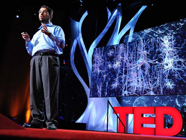
Humans have long held a fascination for the human brain. We chart it, we've described it, we've drawn it, we've mapped it. Now just like the physical maps of our world that have been highly influenced by technology -- think Google Maps, think GPS -- the same thing is happening for brain mapping through transformation.
מזה זמן רב בני-אדם מוקסמים מהמוח. עשינו תרשימים שלו, תארנו אותו, ציירנו אותו, מיפינו אותו. אבל בדיוק כמו מפות העולם שהושפעו רבות מהטכנולוגיה -- לדוגמא מפות גוגל, או GPS -- אותו הדבר קורה היום עם מיפוי מוח בגלל השינויים הרבים.
So let's take a look at the brain. Most people, when they first look at a fresh human brain, they say, "It doesn't look what you're typically looking at when someone shows you a brain." Typically, what you're looking at is a fixed brain. It's gray. And this outer layer, this is the vasculature, which is incredible, around a human brain. This is the blood vessels. 20 percent of the oxygen coming from your lungs, 20 percent of the blood pumped from your heart, is servicing this one organ. That's basically, if you hold two fists together, it's just slightly larger than the two fists.
הבה נעיף מבט על המוח. רוב האנשים, כאשר רואים לראשונה מוח אדם טרי, הם אומרים, "הוא לא נראה כמו מה שבדרך-כלל רואים כאשר מישהו מראה לך מוח." בדרך-כלל, מה שרואים זה מוח שעבר טיפול. הוא אפור. שיכבה חיצונית זו, זוהי מערכת כלי-דם, שהיא מדהימה, סביב המוח האנושי. אלה הם כלי-הדם. 20 אחוז מהחמצן שמגיע מהריאות, 20 אחוז מהדם שנשאב מהלב, משרתים איבר יחיד זה. בגדול, אם מחזיקים שני אגרופים צמודים, הוא קצת יותר גדול מהם.
Scientists, sort of at the end of the 20th century, learned that they could track blood flow to map non-invasively where activity was going on in the human brain. So for example, they can see in the back part of the brain, which is just turning around there. There's the cerebellum; that's keeping you upright right now. It's keeping me standing. It's involved in coordinated movement. On the side here, this is temporal cortex. This is the area where primary auditory processing -- so you're hearing my words, you're sending it up into higher language processing centers. Towards the front of the brain is the place in which all of the more complex thought, decision making -- it's the last to mature in late adulthood. This is where all your decision-making processes are going on. It's the place where you're deciding right now you probably aren't going to order the steak for dinner.
מדענים, בסביבות סוף המאה ה-20, למדו שהם יכולים לעקוב אחר זרם דם כדי למפות בצורה לא-פולשנית, מקומות בהם יש פעילות במוח האדם. לדוגמא, הם יכולים להסתכל בחלק האחורי של המוח, שבדיוק פונים לשם. יש את המוח הקטן; שמחזיק אותנו זקופים ממש עכשיו. הוא מחזיק אותי עומד. הוא קשור בתנועה מתואמת. כאן בצד, זוהי אונה רקתית. זהו המקום בו מתרחש עיבוד השמיעה הראשוני -- כך אתם שומעים את מילותיי, אתם משגרים אותן אל מרכזי עיבוד שפה יותר גבוהים. בקידמת המוח מתרחשים התהליכים היותר מורכבים של מחשבה, קבלת החלטות -- אזור זה הוא האחרון להתפתח בתקופת הבגרות המאוחרת. כאן מתרחשים כל תהליכי קבלת ההחלטות. זה האזור בו אתם מחליטים ברגע זה שאינכם מתכוונים להזמין את הסטייק לארוחה.
So if you take a deeper look at the brain, one of the things, if you look at it in cross-section, what you can see is that you can't really see a whole lot of structure there. But there's actually a lot of structure there. It's cells and it's wires all wired together. So about a hundred years ago, some scientists invented a stain that would stain cells. And that's shown here in the the very light blue. You can see areas where neuronal cell bodies are being stained. And what you can see is it's very non-uniform. You see a lot more structure there. So the outer part of that brain is the neocortex. It's one continuous processing unit, if you will. But you can also see things underneath there as well. And all of these blank areas are the areas in which the wires are running through. They're probably less cell dense. So there's about 86 billion neurons in our brain. And as you can see, they're very non-uniformly distributed. And how they're distributed really contributes to their underlying function. And of course, as I mentioned before, since we can now start to map brain function, we can start to tie these into the individual cells.
כך שאם מתבוננים יותר עמוק לתוך המוח, אם מסתכלים עליו בחתך רוחב, מה שמבחינים הוא שלא ניתן לראות את המבנה לפרטיו. אבל למעשה יש בזה המון פירוט. אלה תאים וחיווטים המחוברים זה לזה. לפני כמאה שנה, כמה מדענים המציאו חומר שצובע תאים. הוא נראה כאן בתכלת בהירה. ניתן לראות אזורים בהם תאי עצב נצבעים. ורואים שזה לא אחיד. יש הרבה מבנים. כך שהחלק החיצוני של המוח הוא הניאו-קורטקס. אם תרצו, זוהי יחידת עיבוד אחת רצופה. אבל ניתן גם לראות דברים מתחת. וכל האזורים הריקים הללו הם האזורים בהם עוברים הסיבים. ככל הנראה הם פחות צפופים בתאים. ישנם כ-86 מיליארד תאי-עצב במוח שלנו. וכפי שרואים, הם אינם מפוזרים באופן אחיד. אופן פיזורם קובע מהותית את צורת פעולתם. וכפי שהזכרתי קודם, מאחר ואנו יכולים עתה להתחיל למפות את פעילות המוח, נוכל להתחיל ולקשור אותם לתאים מוגדרים.
So let's take a deeper look. Let's look at neurons. So as I mentioned, there are 86 billion neurons. There are also these smaller cells as you'll see. These are support cells -- astrocytes glia. And the nerves themselves are the ones who are receiving input. They're storing it, they're processing it. Each neuron is connected via synapses to up to 10,000 other neurons in your brain. And each neuron itself is largely unique. The unique character of both individual neurons and neurons within a collection of the brain are driven by fundamental properties of their underlying biochemistry. These are proteins. They're proteins that are controlling things like ion channel movement. They're controlling who nervous system cells partner up with. And they're controlling basically everything that the nervous system has to do.
הבה נתבונן יותר עמוק. נתבונן בתאי-עצב. כפי שאמרתי, ישנם 86 מיליארד תאי-עצב. יש גם תאים יותר קטנים כפי שרואים. אלה כולם תאים תומכים -- אסטרוציטים, תאי גלייה. העצבים עצמם, הם אלה שמקבלים את הקלט. הם מאחסנים אותו, הם מעבדים אותו. כל עצב מחובר באמצעות סינפסה לעד 10,000 תאי-עצב אחרים במוח שלנו. כל תא-עצב הוא מאוד ייחודי. האופי הייחודי, הן של תאי-העצב הבודדים והן של תאי-העצב בתוך איזור במוח, נקבע על-ידי מאפיינים ביוכימיים בסיסיים. אלה הם חלבונים. חלבונים המווסתים דברים כמו תנועת תעלות יונים. הם שולטים על הקשרים שיוצרים תאי מערכת עצבים. בעיקרון הם שולטים על כל דבר הקשור במערכת עצבים.
So if we zoom in to an even deeper level, all of those proteins are encoded by our genomes. We each have 23 pairs of chromosomes. We get one from mom, one from dad. And on these chromosomes are roughly 25,000 genes. They're encoded in the DNA. And the nature of a given cell driving its underlying biochemistry is dictated by which of these 25,000 genes are turned on and at what level they're turned on.
אם נתמקד ברמה יותר עמוקה, כל החלבונים האלה מקודדים על-ידי הגנומים שלנו. לכל אחד מאיתנו יש 23 זוגות של כרומוזומים. אחד מקבלים מאמא ואחד מאבא. ובכרומוזומים אלה יש בערך 25,000 גנים. הם מקודדים ב-DNA. והאופי של כל תא, שקובע את הביוכימיה שלו, מוכתב על-ידי אילו גנים מתוך ה-25,000 יופעלו ובאיזו רמה הם יופעלו.
And so our project is seeking to look at this readout, understanding which of these 25,000 genes is turned on. So in order to undertake such a project, we obviously need brains. So we sent our lab technician out. We were seeking normal human brains. What we actually start with is a medical examiner's office. This a place where the dead are brought in. We are seeking normal human brains. There's a lot of criteria by which we're selecting these brains. We want to make sure that we have normal humans between the ages of 20 to 60, they died a somewhat natural death with no injury to the brain, no history of psychiatric disease, no drugs on board -- we do a toxicology workup. And we're very careful about the brains that we do take. We're also selecting for brains in which we can get the tissue, we can get consent to take the tissue within 24 hours of time of death. Because what we're trying to measure, the RNA -- which is the readout from our genes -- is very labile, and so we have to move very quickly.
המיזם שלנו שואף להתבונן בפלט הזה, ולהבין איזה מבין 25,000 הגנים הללו מופעלים. לכן מובן מאליו שכדי לבצע מיזם כזה, אנו זקוקים למוחות. לכן שלחנו את טכנאי המעבדה לחפש. חיפשנו מוחות אדם רגילים. התחלנו אצל פתולוג. זה המקום אליו מובאים המתים. אנו מחפשים מוחות אדם רגילים. יש הרבה קריטריונים לפיהם אנו בוחרים מוחות אלה. ברצוננו לוודא שיש בידינו אנשים רגילים בגילאים 20 עד 60, שהם נפטרו במוות טבעי ללא פגיעה מוחית, שאין להם היסטוריה של מחלת נפש, שלא היה שימוש בסמים -- אנו עושים בדיקות רעלים. אנו מאוד נזהרים בנוגע למוחות שאנו בוחרים. כמו-כן אנו בוחרים מוחות שניתן ליטול מהם ריקמה, שאנו יכולים לקבל הסכמה לנטילת הריקמה בתוך 24 שעות משעת הפטירה. מכיוון שהדבר שאנו מנסים למדוד, ה-RNA -- שהוא הפלט מטעם הגנים -- הוא מאוד לא יציב, ולכן עלינו לפעול במהירות רבה.
One side note on the collection of brains: because of the way that we collect, and because we require consent, we actually have a lot more male brains than female brains. Males are much more likely to die an accidental death in the prime of their life. And men are much more likely to have their significant other, spouse, give consent than the other way around.
הערת שוליים על איסוף מוחות: בגלל הדרך בה אנו אוספים, ומשום שאנו זקוקים להסכמה, יש לנו הרבה יותר מוחות של גברים מאשר נשים. לגברים יש סבירות הרבה יותר גבוהה למות באופן לא צפוי בשלב מוקדם של חייהם. ולגברים יש סבירות יותר גבוהה שבת-זוגם תתן את הסכמתה מאשר המצב ההפוך.
(Laughter)
(צחוק)
So the first thing that we do at the site of collection is we collect what's called an MR. This is magnetic resonance imaging -- MRI. It's a standard template by which we're going to hang the rest of this data. So we collect this MR. And you can think of this as our satellite view for our map. The next thing we do is we collect what's called a diffusion tensor imaging. This maps the large cabling in the brain. And again, you can think of this as almost mapping our interstate highways, if you will. The brain is removed from the skull, and then it's sliced into one-centimeter slices. And those are frozen solid, and they're shipped to Seattle. And in Seattle, we take these -- this is a whole human hemisphere -- and we put them into what's basically a glorified meat slicer. There's a blade here that's going to cut across a section of the tissue and transfer it to a microscope slide. We're going to then apply one of those stains to it, and we scan it. And then what we get is our first mapping.
לכן הדבר הראשון שאנו עושים באתר האיסוף זה ליטול את מה שנקרא MR. זוהי הדמיה בתהודה מגנטית -- MRI. זוהי תבנית סטנדרטית שבאמצעותה נציג את כל הנתונים. אנו אוספים את ה-MR. מעין מבט-על לצורך המפה שלנו. הדבר הבא שאנו עושים זו הדמיית DTI. היא ממפה את הכבלים הגדולים במוח. אפשר לחשוב על זה כמעט כמו מיפוי של כבישים מהירים בין-עירוניים. המוח מוסר מהגולגולת, ואז נפרס לפרוסות בנות 1 ס"מ. הן עוברות הקפאה ונשלחות לסיאטל. בסיאטל אנו נוטלים אותן -- זוהי אונת מוח אנושית שלמה -- ואנו שמים אותן בחותך הבשר המהולל. יש להב שיחתוך מקטע מהריקמה, ואז להעבירו אל זכוכית נושאת. לאחר-מכן צובעים אותו ואז סורקים אותו. כך מקבלים את המיפוי הראשון.
So this is where experts come in and they make basic anatomic assignments. You could consider this state boundaries, if you will, those pretty broad outlines. From this, we're able to then fragment that brain into further pieces, which then we can put on a smaller cryostat. And this is just showing this here -- this frozen tissue, and it's being cut. This is 20 microns thin, so this is about a baby hair's width. And remember, it's frozen. And so you can see here, old-fashioned technology of the paintbrush being applied. We take a microscope slide. Then we very carefully melt onto the slide. This will then go onto a robot that's going to apply one of those stains to it. And our anatomists are going to go in and take a deeper look at this.
כאן נכנסים לפעולה המומחים שלנו והם מבצעים משימות אנטומיות פשוטות. אפשר לדמיין את קוי-המיתאר העבים הללו בתור גבולות בין מדינות. מכאן אנו יכולים לפצל את פיסת המוח לחלקים יותר קטנים, שאותם אפשר להניח על-גבי התקן השומר על טמפרטורה נמוכה. כאן פשוט רואים את כל זה -- את הריקמה הקפואה, כאשר היא נחתכת. העובי של זה הוא 20 מיקרון, שזה כמו עובי שיער תינוק. ויש לזכור שזה קפוא. כאן ניתן לראות טכנולוגיה ישנה -- שימוש במברשת צבע. לוקחים זכוכית נושאת ומתיכים מתחתיה בזהירות. אחר-כך זה מועבר לרובוט שיצבע אותו באחד הצבעים ההם. חוקרי האנטומיה יבחנו את זה באופן יותר מעמיק.
So again this is what they can see under the microscope. You can see collections and configurations of large and small cells in clusters and various places. And from there it's routine. They understand where to make these assignments. And they can make basically what's a reference atlas. This is a more detailed map.
וזה מה שהם רואים תחת מיקרוסקופ. ניתן לראות ריכוזים ומבנים של תאים גדולים וקטנים בקבוצות באזורים שונים. מכאן זו עבודה שגרתית. הם יכולים ליצור מין אטלס שהוא מראה-מקום. זו מפה יותר מפורטת.
Our scientists then use this to go back to another piece of that tissue and do what's called laser scanning microdissection. So the technician takes the instructions. They scribe along a place there. And then the laser actually cuts. You can see that blue dot there cutting. And that tissue falls off. You can see on the microscope slide here, that's what's happening in real time. There's a container underneath that's collecting that tissue. We take that tissue, we purify the RNA out of it using some basic technology, and then we put a florescent tag on it. We take that tagged material and we put it on to something called a microarray.
המדענים שלנו משתמשים בזה כדי לחזור לפיסה אחרת מאותה ריקמה ולבצע את מה שקרוי LMD. הטכנאים לוקחים את ההוראות. הם מסמנים לאורך האזור. ואז הלייזר ממש חותך. ניתן לראות את הנקודה הכחולה חותכת. ואותה ריקמה נופלת מטה. ניתן לראות זאת על הזכוכית הנושאת, שזה מה שקורה בזמן אמת. ישנו מיכל למטה אשר קולט את הריקמה. אנו לוקחים את הריקמה, מזקקים ממנה את ה-RNA באמצעות טכנולוגיה פשוטה, ואז מצמידים לה תג פלואורוצנטי. אנו לוקחים את החומר המתוייג ומניחים אותו על-גבי מה שנקרא מערך-מיקרו.
Now this may look like a bunch of dots to you, but each one of these individual dots is actually a unique piece of the human genome that we spotted down on glass. This has roughly 60,000 elements on it, so we repeatedly measure various genes of the 25,000 genes in the genome. And when we take a sample and we hybridize it to it, we get a unique fingerprint, if you will, quantitatively of what genes are turned on in that sample.
זה עשוי להיראות לכם כאוסף של נקודות, אבל כל אחת מהנקודות היא למעשה גן אנושי ייחודי שניקדנו איתו את הזכוכית. יש כאן בערך 60,000 נקודות, וזה אומר שאנו עושים מדידות חוזרות של גנים שונים מ-25,000 הגנים שבגנום. כאשר אנו נוטלים דוגמית ומכליאים אותה עם זה, אנו מקבלים טביעת-אצבע ייחודית כמותית של אילו גנים מופעלים באותה דוגמית.
Now we do this over and over again, this process for any given brain. We're taking over a thousand samples for each brain. This area shown here is an area called the hippocampus. It's involved in learning and memory. And it contributes to about 70 samples of those thousand samples. So each sample gets us about 50,000 data points with repeat measurements, a thousand samples.
אנו עושים זאת שוב ושוב, את התהליך הזה לכל מוח נתון. אנו נוטלים יותר מאלף דוגמיות מכל מוח. אזור זה המוצג כאן נקרא היפוקמפוס. הוא קשור בלמידה וזיכרון. הוא תורם לכ-70 דוגמיות מתוך אלף דוגמיות. לכן כל דוגמית נותנת לנו כ-50,000 נקודות מידע במדידות חוזרות, אלף דוגמיות.
So roughly, we have 50 million data points for a given human brain. We've done right now two human brains-worth of data. We've put all of that together into one thing, and I'll show you what that synthesis looks like. It's basically a large data set of information that's all freely available to any scientist around the world. They don't even have to log in to come use this tool, mine this data, find interesting things out with this. So here's the modalities that we put together. You'll start to recognize these things from what we've collected before. Here's the MR. It provides the framework. There's an operator side on the right that allows you to turn, it allows you to zoom in, it allows you to highlight individual structures.
לכן יש לנו כ- 50 מיליון נקודות מידע למוח אנושי אחד. עד עכשיו הספקנו לאסוף מידע השווה-ערך לשני מוחות אנושיים. חיברנו את הכל ביחד לדבר אחד, ואראה לכם כיצד נראה מיזוג זה. בעיקרון זה מערך גדול של מידע הזמין חינם לכל מדען בעולם. אין צורך אפילו להתחבר כדי להשתמש בכלי זה, לחפור במידע, למצוא עם זה דברים מעניינים. הנה האופנויות שאנו מחברים. תוכלו לזהות דברים אלה ממה שאספנו קודם. הנה ה-MR. הוא מספק את המסגרת. שם מימין יש את הממשק למפעיל שמאפשר להטות, לבצע זום פנימה ולהדגיש מבנים מסויימים.
But most importantly, we're now mapping into this anatomic framework, which is a common framework for people to understand where genes are turned on. So the red levels are where a gene is turned on to a great degree. Green is the sort of cool areas where it's not turned on. And each gene gives us a fingerprint. And remember that we've assayed all the 25,000 genes in the genome and have all of that data available.
אבל הכי חשוב, אנו ממפים היום את המערכת האנטומית הזאת, שהיא המערכת המקובלת כדי שאנשים יבינו היכן הגנים מופעלים. האזורים האדומים הם המקום בו הגן מופעל ברמה גבוהה. ירוק זה מין אזורים קרים בהם הוא אינו מופעל. כל גן נותן לנו טביעת-אצבע. יש לזכור שבחנו את כל 25,000 הגנים שבגנום וכל המידע הזה זמין.
So what can scientists learn about this data? We're just starting to look at this data ourselves. There's some basic things that you would want to understand. Two great examples are drugs, Prozac and Wellbutrin. These are commonly prescribed antidepressants. Now remember, we're assaying genes. Genes send the instructions to make proteins. Proteins are targets for drugs. So drugs bind to proteins and either turn them off, etc. So if you want to understand the action of drugs, you want to understand how they're acting in the ways you want them to, and also in the ways you don't want them to. In the side effect profile, etc., you want to see where those genes are turned on. And for the first time, we can actually do that. We can do that in multiple individuals that we've assayed too.
מה יכולים המדענים ללמוד ממידע זה? אנו בעצמנו רק מתחילים לחקור מידע זה. יש כמה דברים בסיסיים שהיינו רוצים להבין. שתי דוגמאות מצויינות הן של התרופות, פרוזאק וולבוטרין. אלה תרופות שכיחות נגד דיכאון. יש לזכור שאנו בוחנים גנים. גנים שולחים את ההוראות ליצירת חלבונים. חלבונים הם מטרות של תרופות. תרופות מתחברות לחלבונים ובין היתר מפסיקות את פעילותם וכו'. לכן אם רוצים להבין את פעולת התרופות, צריך להבין כיצד הן פועלות בדרכים שאנו רוצים שיפעלו, וגם בדרכים שאין אנו חפצים בהן. בתופעות לוואי וכו', רוצים לראות היכן אותם גנים מופעלים. ולראשונה, אנו ממש יכולים לבצע זאת. אנו גם יכולים לעשות זאת באנשים רבים שבחנו.
So now we can look throughout the brain. We can see this unique fingerprint. And we get confirmation. We get confirmation that, indeed, the gene is turned on -- for something like Prozac, in serotonergic structures, things that are already known be affected -- but we also get to see the whole thing. We also get to see areas that no one has ever looked at before, and we see these genes turned on there. It's as interesting a side effect as it could be. One other thing you can do with such a thing is you can, because it's a pattern matching exercise, because there's unique fingerprint, we can actually scan through the entire genome and find other proteins that show a similar fingerprint. So if you're in drug discovery, for example, you can go through an entire listing of what the genome has on offer to find perhaps better drug targets and optimize.
כעת אנו יכולים להסתכל אל תוך המוח. אנו יכולים לראות את טביעת-האצבע הייחודית. ואנו גם מקבלים אישור לכך. אנו מקבלים אישור שאכן הגן הופעל -- בגלל משהו כמו פרוזאק, במערכות של סרוטונין, אפקט שכבר מוכר לנו -- אבל אנו גם מצליחים לראות את התמונה כולה. אנו גם מצליחים לראות אזורים שאף אחד לא בחן מעולם, ואנו יכולים לראות את הגנים האלה מופעלים שם. זו תופעת לוואי מעניינת. דבר נוסף שניתן לעשות עם זה, בגלל שזה תרגיל בהתאמת תבניות, כי יש טביעת-אצבע ייחודית, אנו יכולים לסרוק את כל הגנום ולמצוא חלבונים אחרים המראים אותה טביעת-אצבע. כך לדוגמא אם רוצים לגלות תרופות חדשות, ניתן לעבור על כל הרשימה של מה שהגנום יכול לתת כדי לאתר אולי מטרות עדיפות עבור התרופות ולהפיק מהן את המירב.
Most of you are probably familiar with genome-wide association studies in the form of people covering in the news saying, "Scientists have recently discovered the gene or genes which affect X." And so these kinds of studies are routinely published by scientists and they're great. They analyze large populations. They look at their entire genomes, and they try to find hot spots of activity that are linked causally to genes. But what you get out of such an exercise is simply a list of genes. It tells you the what, but it doesn't tell you the where. And so it's very important for those researchers that we've created this resource. Now they can come in and they can start to get clues about activity. They can start to look at common pathways -- other things that they simply haven't been able to do before.
רבים מכם ודאי מכירים מחקרי גנום מלא GWAS בצורת התאמות מכלילות של גנום כאשר שדרי חדשות מספרים "מדענים גילו לאחרונה את הגן או הגנים המשפיעים על X". מחקרים מהסוג הזה מתפרסמים כשיגרה על-ידי המדענים וזה טוב. הם מנתחים אוכלוסיות גדולות. הם מסתכלים על הגנום השלם שלהם, ומחפשים אזורי פעילות "חמים" הקשורים באופן סיבתי לגנים. אבל מה שמתקבל ממחקר כזה זו רק רשימה של גנים. היא אומרת לנו מה, אבל לא היכן. לכן זה מאוד חשוב לחוקרים אלה שיצרנו את המאגר הזה. כעת הם יכולים לבוא ולהתחיל לקבל רמזים לגבי פעילות. הם יכולים להסתכל על מסלולים משותפים -- דברים שהם פשוט לא יכלו לעשות קודם.
So I think this audience in particular can understand the importance of individuality. And I think every human, we all have different genetic backgrounds, we all have lived separate lives. But the fact is our genomes are greater than 99 percent similar. We're similar at the genetic level. And what we're finding is actually, even at the brain biochemical level, we are quite similar. And so this shows it's not 99 percent, but it's roughly 90 percent correspondence at a reasonable cutoff, so everything in the cloud is roughly correlated. And then we find some outliers, some things that lie beyond the cloud. And those genes are interesting, but they're very subtle. So I think it's an important message to take home today that even though we celebrate all of our differences, we are quite similar even at the brain level.
אני חושב שקהל זה במיוחד יכול להבין את חשיבות האינדיבידואליות. אני סבור שלכל אדם יש רקע גנטי שונה, כולנו חיינו חיים נפרדים זה מזה. אבל העובדה היא שהגנומים שלנו זהים ביותר מ-99 אחוז. אנו דומים ברמה הגנטית. ומה שאנו מוצאים, שלמעשה, אפילו ברמה הביוכימית של המוח, אנו די דומים. כאן רואים שזה לא 99 אחוז, אלא בקירוב 90 אחוז של התאמה בחתך מייצג, כך שהכל בתוך הענן די תואם אחד לשני. אבל אנו מוצאים כמה מחוץ לתחום, כמה הנמצאים מחוץ לענן. הגנים הללו מעוררים עניין, אבל ההבדלים מאוד עדינים. לכן אני סבור שזה מסר חשוב לקחת היום הביתה והוא שלמרות שאנו משבחים את הנבדלות בינינו, אנו די דומים אפילו ברמת המוח.
Now what do those differences look like? This is an example of a study that we did to follow up and see what exactly those differences were -- and they're quite subtle. These are things where genes are turned on in an individual cell type. These are two genes that we found as good examples. One is called RELN -- it's involved in early developmental cues. DISC1 is a gene that's deleted in schizophrenia. These aren't schizophrenic individuals, but they do show some population variation. And so what you're looking at here in donor one and donor four, which are the exceptions to the other two, that genes are being turned on in a very specific subset of cells. It's this dark purple precipitate within the cell that's telling us a gene is turned on there. Whether or not that's due to an individual's genetic background or their experiences, we don't know. Those kinds of studies require much larger populations.
איך נראים ההבדלים הללו? זוהי דוגמא למחקר שעשינו כדי לעקוב ולראות מה הם בדיוק ההבדלים -- והם די עדינים. אלה הם הדברים בגללם גנים מתעוררים בתאים מסויימים בלבד. אלה הם שני גנים שמצאנו כדוגמאות טובות. אחד נקרא RELN -- קשור באותות התפתחותיים מוקדמים. DISC1 הוא גן שנשמט בסכיזופרניה. אלה לא אנשים סכיזופרניים, אבל הם כן מציגים שוני מהאוכלוסיה הרגילה. אז מה שרואים כאן בתורם 1 ותורם 4, שהם שונים משני האחרים, שגנים מופעלים במערכות-מישנה מאוד מסויימות של תאים. אלה הכתמים הכהים הסגולים בתוך התא האומרים לנו שגן הופעל שם. אם זה בגלל הרקע הגנטי של אותו אדם או בגלל חוויותיו, איננו יודעים. מחקרים כאלה דורשים אוכלוסיות הרבה יותר גדולות.
So I'm going to leave you with a final note about the complexity of the brain and how much more we have to go. I think these resources are incredibly valuable. They give researchers a handle on where to go. But we only looked at a handful of individuals at this point. We're certainly going to be looking at more. I'll just close by saying that the tools are there, and this is truly an unexplored, undiscovered continent. This is the new frontier, if you will. And so for those who are undaunted, but humbled by the complexity of the brain, the future awaits.
לסיום אשאיר אתכם עם הערה אחרונה על מורכבות המוח ועד כמה הרבה עוד עלינו לעבור. אני סבור שמאגרי מידע אלה חשובים עד מאוד. הם נותנים לחוקרים כיוון לאן להתקדם. אבל בדקנו רק קומץ אנשים בשלב זה. בטוח שנבדוק עוד. אסיים באומרי שהכלים כבר קיימים, וזוהי באמת יבשת שלמה שעדיין לא נתגלתה ולא נחקרה. אם תרצו, זוהי החזית החדשה. ולכן עבור כל אלה העשויים ללא חת אבל חשים ענווה מול מורכבות המוח, העתיד מחכה לכם.
Thanks.
תודה.
(Applause)
(מחיאות כפיים)