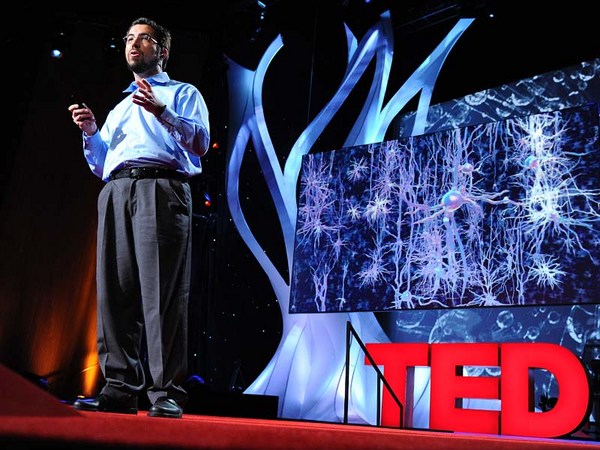
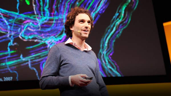
Humans have long held a fascination for the human brain. We chart it, we've described it, we've drawn it, we've mapped it. Now just like the physical maps of our world that have been highly influenced by technology -- think Google Maps, think GPS -- the same thing is happening for brain mapping through transformation.
A los seres humanos siempre nos ha fascinado el cerebro. Lo graficamos, lo describimos, lo dibujamos, lo cartografiamos. Igual que la tecnología afectó a la cartografía, piensen en Google Maps, en el GPS, ocurre la misma transformación en los mapas del cerebro.
So let's take a look at the brain. Most people, when they first look at a fresh human brain, they say, "It doesn't look what you're typically looking at when someone shows you a brain." Typically, what you're looking at is a fixed brain. It's gray. And this outer layer, this is the vasculature, which is incredible, around a human brain. This is the blood vessels. 20 percent of the oxygen coming from your lungs, 20 percent of the blood pumped from your heart, is servicing this one organ. That's basically, if you hold two fists together, it's just slightly larger than the two fists.
Miremos el cerebro. Mucha gente al ver un cerebro real por primera vez dice: "no se parece a lo que se ve generalmente cuando alguien muestra un cerebro". En general se ve un cerebro estático, gris. Esta capa exterior es la vasculatura; algo que recubre al cerebro humano. Estos son los vasos sanguíneos. El cerebro recibe oxígeno en un 20% de los pulmones, el 20% de la sangre que bombea el corazón. Básicamente, si juntamos los dos puños es un poco más grande que los dos puños.
Scientists, sort of at the end of the 20th century, learned that they could track blood flow to map non-invasively where activity was going on in the human brain. So for example, they can see in the back part of the brain, which is just turning around there. There's the cerebellum; that's keeping you upright right now. It's keeping me standing. It's involved in coordinated movement. On the side here, this is temporal cortex. This is the area where primary auditory processing -- so you're hearing my words, you're sending it up into higher language processing centers. Towards the front of the brain is the place in which all of the more complex thought, decision making -- it's the last to mature in late adulthood. This is where all your decision-making processes are going on. It's the place where you're deciding right now you probably aren't going to order the steak for dinner.
A finales del siglo XX los científicos aprendieron a hacer mapas no invasivos siguiendo el flujo sanguíneo, para detectar actividad en el cerebro. Por ejemplo, vean la parte negra del cerebro que gira ahora hacia allí. Ese es el cerebelo; que ahora nos mantiene erguidos. Me mantiene de pie. Participa en los movimientos coordinados. A un lado, aquí tenemos la corteza temporal. Ahí ocurre el procesamiento auditivo primario, por eso oyen mis palabras y las envían a los centros de procesamiento del lenguaje. Hacia la parte frontal del cerebro ocurre el pensamiento más complejo, la toma de decisiones... es lo último que madura en la adultez tardía. Aquí ocurren todos los procesos de toma de decisiones. Es el lugar en el que ahora deciden que no pedirán carne en la cena.
So if you take a deeper look at the brain, one of the things, if you look at it in cross-section, what you can see is that you can't really see a whole lot of structure there. But there's actually a lot of structure there. It's cells and it's wires all wired together. So about a hundred years ago, some scientists invented a stain that would stain cells. And that's shown here in the the very light blue. You can see areas where neuronal cell bodies are being stained. And what you can see is it's very non-uniform. You see a lot more structure there. So the outer part of that brain is the neocortex. It's one continuous processing unit, if you will. But you can also see things underneath there as well. And all of these blank areas are the areas in which the wires are running through. They're probably less cell dense. So there's about 86 billion neurons in our brain. And as you can see, they're very non-uniformly distributed. And how they're distributed really contributes to their underlying function. And of course, as I mentioned before, since we can now start to map brain function, we can start to tie these into the individual cells.
Mirando el cerebro más de cerca, si lo miramos en un corte transversal, podemos advertir que no se ve allí gran estructura. Pero sí hay una gran estructura. Hay células y conexiones, todo está conectado. Hace unos cien años algunos científicos inventaron una tintura celular. Aquí se ve en celeste. Se ven zonas con cuerpos celulares normales coloreados. Como ven es muy irregular. Se ve una gran estructura. La parte exterior del cerebro es el neocórtex. La unidad de procesamiento continuo, si se quiere. Pero ven debajo de eso también. Y en esas zonas blancas están las zonas por las que pasan las conexiones. Quizá haya menor densidad celular. Hay unas 86.000 millones de neuronas en el cerebro. Y, como ven, distribuidas de manera muy irregular. Esta distribución contribuye realmente a sus funciones subyacentes. Y, claro, como dije antes, como ahora podemos mapear la función cerebral podemos llegar a las células individuales.
So let's take a deeper look. Let's look at neurons. So as I mentioned, there are 86 billion neurons. There are also these smaller cells as you'll see. These are support cells -- astrocytes glia. And the nerves themselves are the ones who are receiving input. They're storing it, they're processing it. Each neuron is connected via synapses to up to 10,000 other neurons in your brain. And each neuron itself is largely unique. The unique character of both individual neurons and neurons within a collection of the brain are driven by fundamental properties of their underlying biochemistry. These are proteins. They're proteins that are controlling things like ion channel movement. They're controlling who nervous system cells partner up with. And they're controlling basically everything that the nervous system has to do.
Veamos en más detalle. Veamos las neuronas. Como dije, hay 86.000 millones de neuronas. Además hay células más pequeñas, como verán. Estas son células de apoyo; astrocitos. Y los propios nervios reciben la entrada. La almacenan y la procesan. Cada neurona se conecta por sinapsis hasta con 10.000 neuronas. Y cada neurona, en gran medida, es única. El carácter único de las neuronas individuales y de las neuronas en el conjunto del cerebro sigue las propiedades fundamentales de su bioquímica subyacente. Estas son las proteínas, que controlan el movimiento de los canales iónicos. Controlan qué célula del sistema nervioso se relaciona con otra. Y, básicamente, controlan todo lo que tiene que hacer el sistema nervioso.
So if we zoom in to an even deeper level, all of those proteins are encoded by our genomes. We each have 23 pairs of chromosomes. We get one from mom, one from dad. And on these chromosomes are roughly 25,000 genes. They're encoded in the DNA. And the nature of a given cell driving its underlying biochemistry is dictated by which of these 25,000 genes are turned on and at what level they're turned on.
Así que si nos acercamos aún más todas esas proteínas están codificadas en el genoma. Todos tenemos 23 pares de cromosomas: uno de mamá y uno de papá. Y en estos cromosomas hay unos 25.000 genes. Están codificados en el ADN. Y la naturaleza de una célula, que guía su bioquímica subyacente, depende de cuál de esos 25.000 genes está activado y del nivel de activación.
And so our project is seeking to look at this readout, understanding which of these 25,000 genes is turned on. So in order to undertake such a project, we obviously need brains. So we sent our lab technician out. We were seeking normal human brains. What we actually start with is a medical examiner's office. This a place where the dead are brought in. We are seeking normal human brains. There's a lot of criteria by which we're selecting these brains. We want to make sure that we have normal humans between the ages of 20 to 60, they died a somewhat natural death with no injury to the brain, no history of psychiatric disease, no drugs on board -- we do a toxicology workup. And we're very careful about the brains that we do take. We're also selecting for brains in which we can get the tissue, we can get consent to take the tissue within 24 hours of time of death. Because what we're trying to measure, the RNA -- which is the readout from our genes -- is very labile, and so we have to move very quickly.
Nuestro proyecto intenta observar esta lectura y entender cuáles de estos 25.000 genes están activados. Para realizar un proyecto así obviamente necesitamos cerebros. Mandamos a nuestros técnicos en busca de cerebros normales. Empezamos en la oficina de un médico forense. Allí llevan a los muertos. Buscamos cerebros normales. Seguimos varios criterios al seleccionar cerebros. Queremos asegurarnos que son de humanos normales de entre 20 y 60 años que tuvieron una muerte natural sin lesiones cerebrales ni antecedentes de enfermedad psiquiátrica, ni presencia de drogas. Hacemos análisis toxicológicos. Somos muy cuidadosos con los cerebros que seleccionamos. Otra condición es que podamos obtener el tejido; el consentimiento para tomar el tejido dentro de las 24 horas de la muerte. Porque tratamos de medir el ARN -la lectura de nuestros genes- es muy lábil, por eso tenemos que movernos con rapidez.
One side note on the collection of brains: because of the way that we collect, and because we require consent, we actually have a lot more male brains than female brains. Males are much more likely to die an accidental death in the prime of their life. And men are much more likely to have their significant other, spouse, give consent than the other way around.
Una nota al margen de la selección de cerebros: debido al modo de recolección y dado que hace falta consentimiento, tenemos más cerebros masculinos que femeninos. Los hombres son más propensos a morir en accidentes en la flor de la vida. Y es mucho más probable obtener el consentimiento del cónyuge, la esposa, que al revés.
(Laughter)
(Risas)
So the first thing that we do at the site of collection is we collect what's called an MR. This is magnetic resonance imaging -- MRI. It's a standard template by which we're going to hang the rest of this data. So we collect this MR. And you can think of this as our satellite view for our map. The next thing we do is we collect what's called a diffusion tensor imaging. This maps the large cabling in the brain. And again, you can think of this as almost mapping our interstate highways, if you will. The brain is removed from the skull, and then it's sliced into one-centimeter slices. And those are frozen solid, and they're shipped to Seattle. And in Seattle, we take these -- this is a whole human hemisphere -- and we put them into what's basically a glorified meat slicer. There's a blade here that's going to cut across a section of the tissue and transfer it to a microscope slide. We're going to then apply one of those stains to it, and we scan it. And then what we get is our first mapping.
Lo primero que hacemos en el lugar de recolección es recolectar lo que se llama RM. Es decir, una imagen de resonancia magnética o IRM. Es una plantilla estándar sobre la que estructuraremos el resto de los datos. Así que recolectamos RM. Podemos pensarlo como una vista satelital del mapa. Luego recolectamos imágenes con tensores de difusión: el mapa del gran cableado del cerebro. De nuevo, pueden pensarlo como si cartografiaramos las rutas provinciales. Se extrae el cerebro del cráneo y luego se corta en rodajas de un cm. Todo se congela y se envía a Seattle. Allí, tomamos... esto es un hemisferio completo, y lo pasamos por esta cortadora de carne venida a más. Hay una hoja que corta una sección del tejido y se transfiere al portaobjetos. Luego la coloreamos y la escaneamos. Así construimos nuestro primer mapa.
So this is where experts come in and they make basic anatomic assignments. You could consider this state boundaries, if you will, those pretty broad outlines. From this, we're able to then fragment that brain into further pieces, which then we can put on a smaller cryostat. And this is just showing this here -- this frozen tissue, and it's being cut. This is 20 microns thin, so this is about a baby hair's width. And remember, it's frozen. And so you can see here, old-fashioned technology of the paintbrush being applied. We take a microscope slide. Then we very carefully melt onto the slide. This will then go onto a robot that's going to apply one of those stains to it. And our anatomists are going to go in and take a deeper look at this.
Aquí entran en juego los expertos que realizan tareas básicas de anatomía. Pueden pensarlo como fronteras entre provincias, esas líneas bastante gruesas. A partir de esto podemos fragmentar el cerebro en trozos que luego pondremos en un criostato más pequeño. Aquí mostramos este tejido congelado, que se corta. Tiene 20 micrones de espesor, como el cabello de un bebé. Y recuerden, está congelado. Aquí ven que se usa la vieja tecnología del pincel. Tomamos un portaobjetos. Luego derretimos la lámina cuidadosamente. Después pasa por un robot que lo colorea para que los anatomistas lo observen en detalle.
So again this is what they can see under the microscope. You can see collections and configurations of large and small cells in clusters and various places. And from there it's routine. They understand where to make these assignments. And they can make basically what's a reference atlas. This is a more detailed map.
Esto es lo que se verá bajo el microscopio. Se ven conjuntos y configuraciones de células grandes y pequeñas en grupos y en varios lugares. A partir de ahí es rutina. Ellos saben dónde trabajar. Y construyen un atlas de referencia. Este es un mapa más detallado.
Our scientists then use this to go back to another piece of that tissue and do what's called laser scanning microdissection. So the technician takes the instructions. They scribe along a place there. And then the laser actually cuts. You can see that blue dot there cutting. And that tissue falls off. You can see on the microscope slide here, that's what's happening in real time. There's a container underneath that's collecting that tissue. We take that tissue, we purify the RNA out of it using some basic technology, and then we put a florescent tag on it. We take that tagged material and we put it on to something called a microarray.
Los científicos usan esto para volver a otra pieza de tejido y hacer la microdisección láser. El técnico toma las instrucciones, escribe por allí y luego el láser corta. Allí ven el corte del punto azul. Y ese tejido se desprende. Pueden ver en el portaobjetos lo que sucede en tiempo real. Debajo hay un contenedor que recolecta ese tejido. Tomamos ese tejido purificamos su ARN mediante varias tecnologías básicas y luego le ponemos una etiqueta fluorescente. Tomamos el material etiquetado y lo ponemos en algo llamado micromatriz.
Now this may look like a bunch of dots to you, but each one of these individual dots is actually a unique piece of the human genome that we spotted down on glass. This has roughly 60,000 elements on it, so we repeatedly measure various genes of the 25,000 genes in the genome. And when we take a sample and we hybridize it to it, we get a unique fingerprint, if you will, quantitatively of what genes are turned on in that sample.
Esto parece un puñado de puntos pero cada uno de estos puntos es una pieza única del genoma humano que identificamos en un cristal. Esto contiene unos 60.000 elementos así que medimos muchas veces varios genes de los 25.000 genes del genoma. Y al tomar una muestra e hibridarla con eso obtenemos una huella única, si se quiere, que cuantifica los genes activos en la muestra.
Now we do this over and over again, this process for any given brain. We're taking over a thousand samples for each brain. This area shown here is an area called the hippocampus. It's involved in learning and memory. And it contributes to about 70 samples of those thousand samples. So each sample gets us about 50,000 data points with repeat measurements, a thousand samples.
Hacemos esto una y otra vez para cada cerebro. De cada cerebro tomamos más de mil muestras. Esta zona de aquí es el hipocampo. Se relaciona con el aprendizaje y la memoria y contribuye con cerca de 70 muestras de esas miles. Por eso cada muestra nos da unos 50.000 datos con medidas repetidas, y hay unas mil muestras.
So roughly, we have 50 million data points for a given human brain. We've done right now two human brains-worth of data. We've put all of that together into one thing, and I'll show you what that synthesis looks like. It's basically a large data set of information that's all freely available to any scientist around the world. They don't even have to log in to come use this tool, mine this data, find interesting things out with this. So here's the modalities that we put together. You'll start to recognize these things from what we've collected before. Here's the MR. It provides the framework. There's an operator side on the right that allows you to turn, it allows you to zoom in, it allows you to highlight individual structures.
Hay unos 50 millones de datos para cada cerebro. Hasta ahora tenemos datos equivalentes a dos cerebros. Hemos juntado todo en una sola cosa y les mostraré el aspecto de esa síntesis. Básicamente es un gran conjunto de información disponible gratis para cualquier científico del mundo. Ni siquiera deben registrarse para usar esta herramienta para analizar los datos y encontrar algo interesante. Estas son las modalidades que consolidamos. Empezarán a reconocer cosas a partir de lo ya habíamos recolectado. Esta es la RM que proporciona el marco. Hay un control a la derecha que permite girar, permite aumentar, permite resaltar estructuras individuales.
But most importantly, we're now mapping into this anatomic framework, which is a common framework for people to understand where genes are turned on. So the red levels are where a gene is turned on to a great degree. Green is the sort of cool areas where it's not turned on. And each gene gives us a fingerprint. And remember that we've assayed all the 25,000 genes in the genome and have all of that data available.
Pero, lo más importante, ahora mapeamos en este marco anatómico, un marco común para que se entienda dónde se activan los genes. Los niveles rojos son lugares en los que se activan mucho los genes. Las zonas verdes son más bien frías, no se encienden. Y cada gen deja una huella. Recuerden que analizamos los 25.000 genes del genoma y que tenemos todos los datos disponibles.
So what can scientists learn about this data? We're just starting to look at this data ourselves. There's some basic things that you would want to understand. Two great examples are drugs, Prozac and Wellbutrin. These are commonly prescribed antidepressants. Now remember, we're assaying genes. Genes send the instructions to make proteins. Proteins are targets for drugs. So drugs bind to proteins and either turn them off, etc. So if you want to understand the action of drugs, you want to understand how they're acting in the ways you want them to, and also in the ways you don't want them to. In the side effect profile, etc., you want to see where those genes are turned on. And for the first time, we can actually do that. We can do that in multiple individuals that we've assayed too.
¿Qué pueden aprender los científicos de estos datos? Empezamos a ver estos datos nosotros mismos. Hay cosas básicas que querríamos entender. Dos grandes ejemplos son los medicamentos Prozac y Wellbutrin. Son antidepresivos recetados comúnmente. Recuerden que analizamos genes. Los genes envían instrucciones para crear proteínas. Las proteínas son el objetivo de los medicamentos. Las medicinas se adhieren a las proteínas y las apagan, etc. Si queremos entender el funcionamiento de los medicamentos entender cómo actúan de la forma que queremos, y también de la forma que no queremos. Sus efectos secundarios, etc., queremos ver dónde se activan esos genes. Y, por primera vez, podemos hacerlo. Podemos hacerlo en varias personas analizadas.
So now we can look throughout the brain. We can see this unique fingerprint. And we get confirmation. We get confirmation that, indeed, the gene is turned on -- for something like Prozac, in serotonergic structures, things that are already known be affected -- but we also get to see the whole thing. We also get to see areas that no one has ever looked at before, and we see these genes turned on there. It's as interesting a side effect as it could be. One other thing you can do with such a thing is you can, because it's a pattern matching exercise, because there's unique fingerprint, we can actually scan through the entire genome and find other proteins that show a similar fingerprint. So if you're in drug discovery, for example, you can go through an entire listing of what the genome has on offer to find perhaps better drug targets and optimize.
Ahora miraremos en el cerebro. Podemos ver esta huella única. Y tenemos una confirmación de que, de hecho, el gen se activa con Prozac en estructuras serotoninérgicas, algo de lo que ya se sabe que afecta pero también podemos ver el todo. También vemos zonas que nadie ha visto antes y allí vemos esos genes encendidos. Es muy interesante como efecto secundario. Otra cosa que se puede hacer con algo así dado que es un ejercicio de comparación de patrones, porque hay una huella única, podemos barrer todo el genoma y encontrar otras proteínas que tengan una huella similar. Así que si investigan medicinas, por ejemplo, pueden analizar todo un listado de lo que el genoma ofrece para encontrar quizá mejores objetivos y optimizarlas.
Most of you are probably familiar with genome-wide association studies in the form of people covering in the news saying, "Scientists have recently discovered the gene or genes which affect X." And so these kinds of studies are routinely published by scientists and they're great. They analyze large populations. They look at their entire genomes, and they try to find hot spots of activity that are linked causally to genes. But what you get out of such an exercise is simply a list of genes. It tells you the what, but it doesn't tell you the where. And so it's very important for those researchers that we've created this resource. Now they can come in and they can start to get clues about activity. They can start to look at common pathways -- other things that they simply haven't been able to do before.
A muchos les resultarán familiares los estudios sobre el genoma publicados en las noticias y que dicen: "Los científicos descubrieron el gen o los genes que afectan X". Los científicos publican a menudo este tipo de estudios. Y son geniales. Analizan grandes poblaciones. Miran todos sus genomas y tratan de encontrar zonas de mucha actividad que tengan una relación causal con los genes. Pero de estos ejercicios obtenemos una lista de genes. Se nos dice qué, pero no dónde. Para los investigadores es muy importante que hayamos creado este recurso. Ahora pueden venir y empezar a obtener pistas de esa actividad. Pueden empezar a buscar vías comunes... de otras formas antes inexistentes.
So I think this audience in particular can understand the importance of individuality. And I think every human, we all have different genetic backgrounds, we all have lived separate lives. But the fact is our genomes are greater than 99 percent similar. We're similar at the genetic level. And what we're finding is actually, even at the brain biochemical level, we are quite similar. And so this shows it's not 99 percent, but it's roughly 90 percent correspondence at a reasonable cutoff, so everything in the cloud is roughly correlated. And then we find some outliers, some things that lie beyond the cloud. And those genes are interesting, but they're very subtle. So I think it's an important message to take home today that even though we celebrate all of our differences, we are quite similar even at the brain level.
Creo que esta audiencia en particular entiende la importancia de la individualidad. Pienso que como humanos tenemos diferentes antecedentes genéticos, todos vivimos vidas separadas. Pero el hecho es que son similares en más del 99%. Somos similares a nivel genético. Y hallamos que incluso a nivel de la bioquímica cerebral somos bastante similares. Esto demuestra no el 99%, pero casi el 90% de correspondencia en un corte razonable así que todo en la nube está correlacionado. Luego encontramos valores extremos que están fuera de la nube. Y los genes son interesantes pero muy sutiles. Hay un mensaje importante para llevarse a casa y es que aún si bien celebramos las diferencias somos muy similares incluso a nivel cerebral.
Now what do those differences look like? This is an example of a study that we did to follow up and see what exactly those differences were -- and they're quite subtle. These are things where genes are turned on in an individual cell type. These are two genes that we found as good examples. One is called RELN -- it's involved in early developmental cues. DISC1 is a gene that's deleted in schizophrenia. These aren't schizophrenic individuals, but they do show some population variation. And so what you're looking at here in donor one and donor four, which are the exceptions to the other two, that genes are being turned on in a very specific subset of cells. It's this dark purple precipitate within the cell that's telling us a gene is turned on there. Whether or not that's due to an individual's genetic background or their experiences, we don't know. Those kinds of studies require much larger populations.
¿Cómo son esas diferencias? Este es el ejemplo de un estudio que hicimos para analizar esas diferencias y son muy sutiles. Aquí los genes se encienden en un tipo individual de célula. Estos dos genes son buenos ejemplos. Uno se llama RELN... participa en las claves tempranas del desarrollo. El DISC1 es un gen ausente en la esquizofrenia. Estas no son personas con esquizofrenia pero muestran cierta variación de la población. Y aquí vemos en los donantes uno y cuatro, que son la excepción a los otros dos, esos genes se activan en un subconjunto muy específico de células. Este precipitado de color púrpura oscuro dentro de la célula nos dice que allí hay un gen activo. Se deba o no a los antecedentes genéticos de una persona o a sus experiencias no lo sabemos. Esos estudios requieren de poblaciones mucho más grandes.
So I'm going to leave you with a final note about the complexity of the brain and how much more we have to go. I think these resources are incredibly valuable. They give researchers a handle on where to go. But we only looked at a handful of individuals at this point. We're certainly going to be looking at more. I'll just close by saying that the tools are there, and this is truly an unexplored, undiscovered continent. This is the new frontier, if you will. And so for those who are undaunted, but humbled by the complexity of the brain, the future awaits.
Terminaré con una nota final sobre la complejidad del cerebro y lo que falta por recorrer. Creo que estos recursos son muy valiosos. Son una guía para investigadores. Pero en este momento sólo vimos un puñado de personas. Sin duda incorporaremos más. Terminaré diciendo que las herramientas existen, que es un continente inexplorado, sin descubrir. Es la nueva frontera, si se quiere. Y para los intrépidos, intimidados por la complejidad del cerebro, el futuro aguarda.
Thanks.
Gracias.
(Applause)
(Aplausos)